
Coronavirus Testing in the Outpatient Setting - CAM 380
Description
Human coronaviruses, first characterized in the 1960s, are named based on the spiked proteins located on their surface. As of 2020, seven coronaviruses are known to infect humans. Four, of which—229E, NL63, OC43, and HKU1—are associated with the common cold. MERS-CoV is the coronavirus that causes Middle East Respiratory Syndrome, or MERS. SARS-CoV is the causative agent of Severe Acute Respiratory Syndrome (SARS), and SARS-CoV-2 is the virus that causes coronavirus disease 2019, or COVID-19.1 As of June 1, 2024, the United States had reported that nearly 1.2 million people have died of COVID-19.1 Testing for a possible coronavirus infection can include molecular tests, such as nucleic acid-based testing like reverse transcription polymerase chain reaction (RT-PCR); host antibody testing; and antigen testing.
Policy
Application of coverage criteria is dependent upon an individual’s benefit coverage at the time of the request:
- Targeted nucleic acid testing (e.g., RT-PCR, rapid molecular tests) for COVID-19 (SARS-CoV-2) is considered MEDICALLY NECESSARY in any of the following situations:
- For individuals displaying signs and symptoms of possible COVID-19 infection (See Note 1).
- For asymptomatic individuals with known exposure to COVID-19, EXCEPT when the individual has had a previous COVID-19 infection within the last 90 days.
- For individuals with signs or symptoms of SARS and who have traveled to endemic areas or who have been exposed to persons with SARS, targeted nucleic acid testing (e.g., RT-PCR) for the detection of severe acute respiratory syndrome (SARS) coronavirus RNA is considered MEDICALLY NECESSARY.
- For individuals with signs or symptoms of Middle East respiratory syndrome (MERS) and who have traveled to endemic areas or who have been exposed to persons with MERS, targeted nucleic acid testing (e.g, RT-PCR) for the detection of MERS coronavirus RNA is considered MEDICALLY NECESSARY.
- To support a diagnosis of multisystem inflammatory syndrome in children (MIS-C) (see Note 2), multisystem inflammatory syndrome in adults (MIS-A) (see Note 3), or post-acute sequelae of SARS-CoV-2 infection (PASC), nucleic acid amplification testing and host antibody serology testing is considered MEDICALLY NECESSARY.
- For symptomatic individuals, antigen-detecting diagnostic tests for SARS-CoV-2 (e.g., antigen rapid tests) once every 48 hours is considered MEDICALLY NECESSARY.
- For the diagnosis of SARS-CoV-2 reinfection, whole genome sequencing of paired specimens from distinct lineages (as defined in Nextstrain or GISAID) is considered NOT MEDICALLY NECESSARY.
- For all other situations not described above, host antibody serology testing is considered NOT MEDICALLY NECESSARY.
The following does not meet coverage criteria due to a lack of available published scientific literature confirming that the test(s) is/are required and beneficial for the diagnosis and treatment of an individual’s illness.
- In the outpatient setting, SARS-CoV-2 genotyping is considered NOT MEDICALLY NECESSARY.
- For all situations, neutralization antibody testing for SARS-CoV-2 is considered NOT MEDICALLY NECESSARY.
- Testing for other endemic coronaviruses, such as 229E, NL63, OC43, and HKU1, is considered NOT MEDICALLY NECESSARY.
NOTES:
Note 1: Signs and symptoms associated with a possible COVID-19 infection can include fever, cough, fatigue, shortness of breath or difficulty breathing, congestion or runny nose, chills, muscle or body aches, headache, sore throat, new loss of taste or smell, nausea, vomiting, and diarrhea.3
Note 2: According the CDC,4 MIS-C is defined as an illness that is found in a person less than 21 years of age when all of the following conditions are met:
- Subjective or documented fever of at least 38°C;
- Clinical severity requiring hospitalization;
- Evidence of systemic inflammation indicated by C-reactive protein (CRP) ≥3.0 mg/dL (30 mg/L);
- New onset of manifestations in at least two of the following categories:
- Cardiac involvement indicated by one of the following:
- Left ventricular ejection fraction <55%.
- Coronary artery dilatation, aneurysm, or ectasia.
- Elevated troponin.
- Mucocutaneous involvement indicated by one of the following:
- Rash.
- Inflammation of the oral mucosa.
- Conjunctivitis or conjunctival injection.
- Extremity findings (e.g., erythema or edema of the hands or feet).
- Shock.
- Gastrointestinal involvement indicated by one of the following:
- Abdominal pain.
- Vomiting.
- Diarrhea.
- Hematologic involvement indicated by one of the following:
- Platelet count <150,000 cells/µL.
- Absolute lymphocyte count (ALC) <1,000 cells/μL.
- Cardiac involvement indicated by one of the following:
Note 3: According to the CDC,3 MIS-A is defined as an illness that is found in a person 21 years of age or older when all of the following conditions are met:
- Hospitalization for 24 hours or more;
- Subjective or documented fever of at least 38°C for one of the following:
- 24 or more hours prior to hospitalization.
- Within the first 3 days of hospitalization.
- No alternative diagnosis (e.g., bacterial sepsis, exacerbation of a chronic medical condition).
- At least three of the following (occurring prior to hospitalization or within the first three days of hospitalization), with at least one being a primary clinical criterion:
- Primary clinical criteria:
- Severe cardiac illness (e.g., myocarditis, pericarditis, coronary artery dilation/aneurysm, new-onset right or left ventricular dysfunction, 2nd/3rd degree A-V block, ventricular tachycardia).
- Rash and non-purulent conjunctivitis.
- Secondary clinical criteria:
- New-onset neurologic signs and symptoms (e.g., encephalopathy in an individuals without prior cognitive impairment, seizures, meningeal signs, peripheral neuropathy including Guillain-Barré syndrome).
- Shock or hypotension not attributable to medical therapy.
- Abdominal pain, vomiting, or diarrhea.
- Thrombocytopenia (platelet count <150,000/μL).
- Primary clinical criteria:
- Evidence of SARS-CoV-2 infection;
- Evidence of systemic inflammation (elevated CRP, ferritin, interleukin-6, erythrocyte sedimentation rate, or procalcitonin).
Table of Terminology
| Term |
Definition |
| 2019-nCoV |
2019 novel coronavirus |
| AACC |
American Association for Clinical Chemistry |
| AAP |
American Academy of Pediatrics |
| ACE-2 |
Angiotensin converting enzyme-2 |
| ACR |
American College of Rheumatology |
| ACS |
American Chemical Society |
| Ag-RDTs |
Antigen-detecting rapid diagnostic tests |
| AMA |
American Medical Association |
| APSF |
Anesthesia Patient Safety Foundation |
| ARDS |
Acute respiratory distress syndrome |
| ASA |
American Society of Anesthesiologists |
| ASM |
American Society for Microbiology |
| BAL |
Bronchoalveolar lavage |
| BAW |
Bulk acoustic wave (biosensors) |
| BNP |
B-type natriuretic peptide |
| CARES Act |
Coronavirus Aid, Relief, And Economic Security Act |
| Cas12a |
CRISPR associated protein 12a |
| CBC |
Complete blood cell count |
| CDC |
Centers For Disease Control and Prevention |
| cDNA |
Complementary deoxyribonucleic acid |
| CFR |
Code of Federal Regulations |
| CI |
Confidence interval |
| CLIA |
Chemiluminescence enzyme immunoassay |
| CLIA ’88 |
Clinical Laboratory Improvement Amendments Of 1988 |
| CMS |
Centers For Medicare & Medicaid Services |
| COVID-19 |
Coronavirus disease 2019 |
| CPK |
Creatine phosphokinase |
| CRP |
C-reactive protein |
| CSSE |
Center for Systems Science and Engineering |
| CT |
Cycle threshold |
| cVNT |
Competitive neutralization test |
| DNA |
Deoxyribonucleic acid |
| DPP7 |
Dipeptidyl peptidase 7 |
| ECDC |
European Centre for Disease Prevention and Control |
| ESR |
Erythrocyte sedimentation rate |
| ETS |
Emergency temporary standard |
| EU/EEA |
European Union / European Economic Area |
| EUA |
Emergency use authorization |
| FAQ |
Frequently asked questions |
| FDA |
Food and Drug Administration |
| FET |
Field-effect transistor |
| FIA |
Fluorescence immunoassays |
| Flu SC2 |
Influenza SARS-CoV-2 (multiplex assay) |
| FN |
False negative |
| FP |
False positive |
| GISAID |
Global initiative on sharing all influenza data |
| GOLGA3 |
Golgi autoantigen, golgin subfamily a, 3 |
| GRADE |
Grading Of Recommendations Assessment, Development, and Evaluation |
| HCoV |
Human coronavirus |
| HCP |
Health care personnel |
| HCW |
Healthcare worker |
| HHS |
Health And Human Services |
| HKU1 |
Human coronavirus |
| HLA |
Human leukocyte antigen |
| HSCT |
Hematopoietic stem cell transplant |
| IMCA |
Immunochemiluminometric assay |
| ICR |
Investigative criteria for suspected cases of SARS-CoV-2 reinfection |
| IDSA |
Infectious Diseases Society of America |
| IFU |
Instructions for use |
| IgA |
Immunoglobulin A |
| IgG |
Immunoglobulin G |
| IgM |
Immunoglobulin M |
| IL-1 |
Interleukin 1 |
| IL-6 |
Interleukin 6 |
| INR |
International normalized ratio |
| IQR |
Interquartile range |
| IVIG |
Intravenous immunoglobulin |
| JAMA |
Journal of the American Medical Association |
| LDH |
Lactic acid dehydrogenase |
| LDTs |
Laboratory-developed tests |
| LFIAs |
Lateral flow immunoassays |
| LoD |
Limit of detection |
| MERS |
Middle east respiratory syndrome |
| MERS-CoV |
Middle east respiratory syndrome–related coronavirus |
| MESIA |
Magnetic force-assisted electrochemical sandwich immunoassay |
| MHRA |
Medicines & Healthcare Products Regulatory Agency |
| MIS-A |
Multisystem inflammatory syndrome in adults |
| MIS-C |
Multisystem inflammatory syndrome in children |
| MMWR |
Morbidity And Mortality Weekly Report |
| mRNA |
Messenger RNA |
| MT |
Mid-turbinate |
| N |
Nucleocapsid |
| NAAT |
Nucleic acid amplification test |
| NAb |
Neutralizing antibody |
| NGS |
Next-generation sequencing |
| NIH |
National Institutes of Health |
| NP |
Nasopharyngeal |
| NPA |
Negative percent agreement |
| NT-proBNP |
N-terminal pro hormone BNP |
| NW |
Nasopharyngeal wash/aspirate or nasal wash/aspirate |
| OD |
Optical density |
| OP |
Oropharyngeal |
| opvCRISPR |
One-pot visual SARS-CoV-2 detection system |
| OSHA |
Occupational Safety and Health Administration |
| PASC |
Post-Acute Sequelae Of SARS-CoV-2 Infection |
| PCR |
Polymerase chain reaction |
| PEM |
Post-exertional malaise |
| PHE |
Public Health England |
| PHS Act |
Public Health Service Act |
| POC |
Point-of-care |
| POC/NP |
Point-of-care/near person |
| PPA |
Positive percent agreement |
| PPE |
Personal protective equipment |
| pro-BNP |
Pro hormone B-type natriuretic peptide |
| PSO |
Past symptom onset |
| PT |
Prothrombin time |
| PTT |
Partial thromboplastin time |
| ptxP |
Single-copy promoter target |
| RADT |
Rapid antigen detection test |
| RBD |
Receptor binding domain |
| RdRp |
Ribonucleic acid-dependent ribonucleic acid polymerase |
| RNA |
Ribonucleic acid |
| RP |
Ribonuclease P gene |
| RP |
Respiratory pathogen |
| RP2 |
Respiratory panel 2 |
| RP2.1 |
Respiratory panel 2.1 |
| RSV |
Respiratory syncytial virus |
| RT |
Reverse transcriptase |
| RT-LAMP |
Reverse transcription loop-mediated isothermal amplification |
| RT-PCR |
Reverse transcription polymerase chain reaction |
| RT-qPCR |
Reverse transcription quantitative PCR |
| SARS-CoV |
Severe acute respiratory syndrome- coronavirus |
| SARS-CoV-2 |
Severe acute respiratory syndrome coronavirus 2 |
| SHEA |
Society for Healthcare Epidemiology of America |
| SNP |
Single nucleotide polymorphism |
| SOT |
Solid organ transplant |
| ssDNA |
Single-stranded deoxyribonucleic acid |
| sVNT |
Surrogate viral neutralization test |
| TCID50 |
Median tissue culture infective dose |
| TMA |
Transcription-mediated amplification |
| TMEM189–UBE2V1 |
PEDS1-UBE2V1 readthrough |
| TN |
True negative |
| TP |
True positive |
| UCSD |
University of California San Diego |
| UTM-RT |
Universal Transport Medium (RT context) |
| VBM |
Variants being monitored |
| VOC |
Variant of concern |
| VOHC |
Variants of high consequence |
| VOI |
Variants of interest |
| WGS |
Whole genome sequencing |
| WHO |
World Health Organization |
Reimbursement
- AMA standard practice for COVID-19 testing states not to include both the HCPCS and AMA code for the same procedure on the same DOS and that only one code should be used, therefore only one code per date of service will be reimbursed.
- Specimen collection codes for coronavirus testing are considered incidental and will not be reimbursed
Rationale
On March 11, 2020, the World Health Organization (WHO) declared the novel coronavirus SARS-CoV-2, or COVID-19, a global pandemic.4 COVID-19 is the third recent human coronavirus to be declared an emergency. SARS (Severe Acute Respiratory Syndrome) was recognized as an emergency by the WHO in February 2003.5 This outbreak in 2003 resulted in over 8000 cases in 26 different countries. Since 2003, only four limited reoccurrences have been reported according to the WHO—three incidences are due to laboratory accidents (in Taipei and Singapore) and one incident of undetermined source in China.5 As early as September 2012, another human coronavirus, MERS-CoV, began to spread in the Middle East, causing Middle East Respiratory Syndrome (MERS). Although the WHO did not initially declare MERS an emergency, they have since added MERS to their list of pandemic/epidemic diseases. Since September 2012 and as of the end of October 2021, the WHO reports 2574 laboratory-confirmed cases of MERS with 858 MERS-associated deaths (34.4% fatality rate) in 27 countries.6
Unlike the initial SARS and MERS outbreaks that were predominantly regionally contained, COVID-19 became a global pandemic. WHO has noted that SARS-CoV-2 continues to cause acute disease and post-COVID-19 condition, although the overall global public health risk was assessed as moderate as of mid-2025.7 Infection from the novel human coronavirus SARS-CoV-2 can result in coronavirus disease 2019 (COVID-19). “Infection with SARS-CoV-2 can range from asymptomatic or mild illness to severe disease with respiratory failure, multiorgan dysfunction, and death, with older adults and individuals with underlying medical conditions at greater risk for poor outcomes.”7 Older individuals and patients with comorbidities—such as cardiovascular disease, diabetes mellitus, hypertension, chronic lung disease, cancer, chronic kidney disease, obesity, and smoking—have an increased likelihood of poor outcomes.8 Sepsis, multiorgan failure (including the kidney, liver, and heart), pneumonia, and acute respiratory distress syndrome (ARDS) can also occur.9,10 Severe outcomes have been associated with the following laboratory features: lymphopenia, elevated liver enzymes, elevated lactate dehydrogenase (LDH), elevated inflammatory markers (such as CRP and ferritin), elevated D-dimer, elevated prothrombin time (PT), elevated troponin, elevated creatine phosphokinase (CPK), and acute kidney injury.8
The global spread of SARs-CoV-2 has been attributed to its high transmissibility compared to other coronaviruses, such as SARS-CoV and MERS-CoV, which is influenced in part by viral load and shedding dynamics. Viral load refers to the quantity of virus present in a given biological sample at a specific point in time and, for SARS-CoV-2, is most commonly measured in respiratory specimens, where viral replication primarily occurs. The viral load of SARS-CoV-2 “peaks around the time of symptom onset, followed by a gradual decrease to a low level after about 10 days. Regarding the period of high infectiousness, a recent study reported that exposure to an index case within five days of symptom onset confers a high risk of secondary transmission.”11 This finding was corroborated by other studies, which found that “SARS-CoV-2 viral load in the upper respiratory tract appeared to peak in the first week of illness, whereas that of SARS-CoV peaked at days 10–14 and that of MERS-CoV peaked at days 7–10;” because SARS-CoV-2 viral load peaks faster, it can be more transmissible earlier in the disease course.12 However, after reaching its peak during symptom onset, the viral load decreases “monotonically.”11 If viral loads do not decrease, patients will be more likely to suffer worse outcomes and require hospitalization.13 Viral load has been found to be either similar among symptomatic and asymptomatic COVID-19 positive individuals, or higher among symptomatic individuals.11 Infectiousness of COVID-19 also correlates with shedding, meaning that the viral particles can replicate in an individual and spread in the environment to others. The mean duration of SARS-CoV-2 RNA shedding “was 17.0 days (95% CI 15·5–18·6; 43 studies, 3229 individuals) in upper respiratory tract, 14.6 days (9·3–20·0; seven studies, 260 individuals) in lower respiratory tract, 17.2 days (14·4–20·1; 13 studies, 586 individuals) in stool, and 16.6 days (3·6–29·7; two studies, 108 individuals) in serum samples,” with maximum shedding duration reaching “83 days in the upper respiratory tract, 59 days in the lower respiratory tract, 126 days in stools, and 60 days in serum.”12
In children and adolescents, reports of a multisystem inflammatory syndrome (MIS-C) with similarities to Kawasaki disease and toxic shock syndrome have been linked to COVID-19.14-17 Multisystem inflammatory syndrome has also been reported in adults (MIS-A). From June to October 2020, researchers reported 27 cases of MIS-A in the US and UK.18 The case definition of MIS-A includes“(1) hospitalization without evidence of severe respiratory illness (to exclude hypoxia as the cause of the signs and symptoms), (2) extrapulmonary organ system involvement (including hypotension or shock, cardiac dysfunction, arterial or venous thromboembolism, acute liver injury, or dermatologic abnormalities), and (3) laboratory evidence of acute inflammation (e.g., highly elevated C-reactive protein, ferritin, D-dimer, or interleukin-6).”18 Most patients present with a fever >100.4 °F, cardiac abnormalities (arrhythmias, elevated troponin levels, or left or right ventricular dysfunction), and gastrointestinal symptoms. Rare symptoms include dermatological manifestations or respiratory symptoms such as pleural effusion. Patients may have elevated laboratory markers of inflammation including CRP, ferritin, and markers of coagulopathy including D-dimer.19
As SARS-CoV-2 has continued to evolve, public health agencies have updated how variants are monitored and classified. Earlier in the pandemic, the CDC categorized variants into multiple groups, including variants being monitored (VBM), variants of interest (VOI), variants of concern (VOC), and variants of high consequence (VOHC), based on characteristics such as transmissibility, disease severity, and impact on diagnostics, therapeutics, and immunity.20 The CDC has since archived this classification framework and associated guidance and in current practice, variant tracking has shifted to ongoing genomic surveillance and lineage-based monitoring rather than reliance on formal categorical designations. Most circulating SARS-CoV-2 variants are now descendants of the Omicron lineage (B.1.1.529), and public health efforts focus on identifying emerging sublineages with potential changes in transmissibility, immune escape, or clinical impact. While variant classification frameworks remain useful for describing potential risk, ongoing surveillance continues to inform public health guidance, vaccine updates, and therapeutic strategies.21
The CDC indicates three vaccines as authorized and recommended to prevent COVID-19 in the US: Pfizer-BioNTech COVID-19 Vaccine, Bivalent; Moderna COVID-19 Vaccine, Bivalent; and Novavax COVID-19 Vaccine, Adjuvanted. The Pfizer-BioNTech and Moderna COVID-19 vaccines are mRNA vaccines, which instruct B and T lymphocytes to fight off that specific mRNA-encoded protein from COVID-19 in the event of future exposure. Novavax is a protein subunit vaccine that delivers pieces (spike proteins) of the virus that causes COVID-19, as well as an adjuvant that helps the immune system respond in the event of future exposure.22
Besides the viruses associated with SARS, MERS, and COVID-19, four other human coronaviruses (HCoVs) are currently known—229E, NL63, OC43, and HKU1. These four viruses are considered endemic to the human population, and they typically cause mild respiratory tract infections associated with the common cold; in fact, it is approximated that up to one-third of all “common colds” may be due to one of these four endemic human coronaviruses. These HCoVs can cause both upper and lower respiratory infections, but they typically result in relatively mild, or even asymptomatic, cases. In immunosuppressed individuals, including those with pre-existing pulmonary diseases, progression to acute respiratory failure can occur in some cases.23,24
Nucleic Acid Testing for Human Coronavirus Infections
Coronaviruses are a family of enveloped, single-stranded positive-sense RNA viruses. During the initial phase of infection, viral RNA is present in respiratory specimens and can be detected using nucleic acid amplification tests (NAATs), such as reverse transcription polymerase chain reaction (RT-PCR). These tests detect viral genetic material and are the preferred method for diagnosing acute SARS-CoV-2 infection. Viral RNA levels are typically highest near the time of symptom onset and decline over the course of infection, although detectable RNA may persist for variable periods following clinical recovery.25 RT-PCR results of SARS-CoV-2 may fluctuate and become unstable over time. Advances in testing technology have enabled both laboratory-based and rapid molecular platforms, allowing for sensitive detection of SARS-CoV-2 in a variety of clinical settings, including point-of-care. While RT-PCR remains highly sensitive, test performance depends on factors such as timing of specimen collection, specimen type, and sample quality.26
Clinical Utility and Validity of Nucleic Acid Testing
Many studies have been performed to date to evaluate the analytical performance of RT-PCR, noting they are analytically sensitize and standardized The WHO-recommended diagnostic RT-PCR protocol targets two genes--the E gene as the molecular target (where the limit is 3.9 copies per reaction) and the RdRp gene as the molecular target (limit of 3.6 copies per reaction).27 One study reported possible in vitro cross-reactivity between the RdRp-based method used predominantly in European labs with SARS-CoV in cell culture.28 SARS-CoV is the coronavirus that caused the initial SARS (Severe Acute Respiratory Syndrome) outbreak in 2003.5 The likelihood of either a co-infection of SARS-CoV and SARS-CoV-2 or a concurrent outbreak of both viruses is small.
The CDC RT-PCR Diagnostic Panel detects SARS-CoV-2 virus in the upper and lower respiratory specimen is an early example of a NAAT test for detecting SARS-CoV-2.29 The CDC diagnostic panel test does not target the RdRp gene; it consists of two primer/probe sets of the N gene and one primer/probe set for human RNase P gene (RP) as the control. The CDC diagnostic panel has a reported limit of 1.0 – 3.2 copies/µL.27 Reports of initial negative RT-PCR results in individuals who later develop symptomatic COVID-19 have been published, it is important to remember that “Negative results do not preclude SARS-CoV-2 infection and should not be used as the sole basis for patient management decisions. Negative results must be combined with clinical observations, patient history, and epidemiological information.”30
Host Antibody Testing
The COVID-19 illness begins with an initial infection by SARS-CoV-2 characterized by the production of antibodies, such as IgM, IgA, and IgG. However, there is a delay between the time of initial infection and the production of immunoglobulins with most individuals seroconverting within one to three weeks following infection.31,32 A study by Kaduskar, et al. (2022) found that IgM and IgG antibodies may appear concurrently or sequentially, and the timing and magnitude of the response vary by individual and clinical factors.33
Serological host antibody tests can detect the presence of IgM and IgG antibodies that an individual has developed in response to an infection—in this case, a SARS-CoV-2 viral infection. The test may report total antibodies present, meaning either it does not distinguish between IgG and IgM or that it is reporting the sum of IgG and IgM. This is sometimes referred to as “total antibody testing.” On the other hand, the test may be specific for one antibody, such as IgG or IgM, or the test may claim to accurately distinguish between the antibodies. Serology testing is currently used for determining past infection and not for diagnostic purposes.
Another type of antibody testing is “neutralizing” antibody detection, as opposed to “binding” antibody detection described above. This process involves incubating serum with a live version of the virus. The analytes of interest are the antibodies that have the capability to prevent infection by the virus (i.e., neutralization). Identification of these antibodies may contain useful clinical information and are often reported in an aggregate titer, as opposed to specifying each individual antibody.34
Interpretation of serologic results can be complicated by widespread vaccination and prior infection, as antibody presence does not reliably distinguish between these sources of immunity and may reflect combined or hybrid immune responses.35
Clinical Utility and Validity of Host Antibody Testing
Antibody testing has many potential uses. Ideally, the use of an accurate, reliable antibody test could possibly show whether someone has previously been exposed to the virus. This could indicate possible immunity in an individual. Please note that the antibody test is not used as a diagnostic test, meaning it should not be used to diagnose an acute infection. Within the FDA policy for diagnostic testing for COVID-19, issued on November 15, 2021 they state, “Results from antibody testing should not be used to diagnose or exclude SARS-CoV-2 infection.”36
The timing of seroconversion following SARS-CoV-2 infection is better characterized as of 2026. Most individuals develop detectable antibodies within 1–3 weeks after symptom onset, with median seroconversion occurring around 10–14 days.31,37 IgM and IgG antibodies may appear concurrently or sequentially, and nearly all infected individuals demonstrate seropositivity within three weeks. However, the exact timing and magnitude of antibody responses vary depending on factors such as disease severity, host immune status, and assay characteristics.31 Antibody levels, particularly IgG, may persist for months following infection, although titers typically decline over time.38 Serologic testing has been recommended for checking prior exposure or for epidemiologic studies rather than for diagnosing acute infection, where molecular or antigen-based testing is preferred.25 Ideally, rapid diagnostic testing in the outpatient setting must be accurate and reliable. Current evidence and public health guidance indicate that viral tests (nucleic acid amplification tests [NAATs] or antigen tests) are the preferred methods for diagnosing acute SARS-CoV-2 infection, as they directly detect the presence of the virus.25
Early in the COVID-19 pandemic, a meta-analysis studies by Lisboa Bastos, et al. (2020) and Kontou, et al. (2020) found substantial variability in the diagnostic accuracy of serologic assays, with lower sensitivity observed in the first week following symptom onset and significant risk of bias across included studies. The authors concluded that t available evidence at the time did not support the use of point-of-care serologic tests for diagnosis.39,40 These findings reflect early-pandemic limitations in assay performance and study quality. Since that time, the role of serologic testing has been more clearly defined, and current guidelines no longer recommend antibody testing for the diagnosis of acute SARS-CoV-2 infection.
Caturegli, et al. (2020) performed a case-control study to determine the clinical utility and validity of using SARS-CoV-2 antibodies, which were serum IgG and IgA antibodies formed against the SARS-CoV-2 spike protein detected by enzyme-linked immunosorbent assay (ELISA). When assays were formed 14 days or later after symptom onset, the researchers found that the sensitivity was 0.976 (95% CI, 0.928 to 0.995) and specificity was 0.988 (95% CI, 0.974 to 0.995), but the sensitivity decreased at earlier time points. Antibodies “predicted the odds of developing acute respiratory distress syndrome, which increased by 62% (CI, 48% to 81%; P < 0.001) for every 2-fold increase in IgG.” This demonstrates the linkage of antibodies used to measure clinical severity and for those who tested negative by NAAT but remained potentially COVID-positive.
Fox, et al. (2022) performed a meta-analysis to assess the accuracy of antibody tests. The analysis covered 178 studies with a total of 64,688 samples taken from 25,724 people with confirmed SARS-CoV-2. All the studies were conducted before the introduction of the SARS-CoV-2 vaccines to ensure the responses were due to naturally acquired antibodies. The average sensitivity for either IgG or IgG combined with IgM was 41.1% one week after symptom onset, 74.9% two weeks after symptom onset, and 88.0% three weeks after symptom onset. The average sensitivity during the convalescent phase of infection, up to 100 days since symptom onset, was 89.8% for IgG, 92.9% for IgG or IgM combined, and 94.3% for total antibodies. The average sensitivities for IgM alone “followed a similar pattern but were of a lower test accuracy in every time slot.” The authors conclude that antibody tests “could be a useful diagnostic tool” but note that “antibody tests have an increasing likelihood of detecting an immune response to infection as time since onset of infection progresses and have demonstrated adequate performance for detection of prior infection for sero-epidemiological purposes” and “the applicability of results for detection of vaccination-induced antibodies is uncertain.”42
Antigen Testing
Another possible diagnostic testing methodology is antigen detection testing, which relies upon the direct detection of parts of the virus called “antigens”—in this instance, proteins located on the outside of SARS-CoV-2, such as the spike protein (S) or nucleocapsid (N) protein, that can cause an immune response in an individual. What makes this method of testing distinct from antibody testing is that antigen testing directly measures the presence of the virus in a person whereas antibody testing is measuring the patient’s response to an infection. These tests are widely available in point-of-care and at-home formats and typically provide results within 15–30 minutes.43
Several antigen testing platforms initially received Emergency Use Authorization (EUA) early in the COVID-19 pandemic and remain in use today. For example, the BD Veritor™ System for SARS-CoV-2, a rapid chromatographic immunoassay, has transitioned from EUA to FDA 510(k) clearance and continues to be used in point-of-care settings.44 Similarly, the LumiraDx SARS-CoV-2 Antigen Test is an automated immunofluorescence assay that provides rapid results as fast as 12 minutes and has been authorized under an EUA.45 Additional platforms, including the Quidel Sofia® antigen test system (now succeeded by the Sofia 2 Flu + SARS Antigen FIA), were among the first antigen tests authorized for SARS-CoV-2 detection and contributed to the expansion of rapid testing capacity.46
A current list of FDA-authorized SARS-CoV-2 diagnostic and multiplex assays is maintained through the NIH Rapid Acceleration of Diagnostics (RADx®) program.43 These testing methods include (among others): Bulk Acoustic Wave (BAW) Biosensors, Chemiluminescence Immunoassays, Chromatographic Digital Immunoassays, Digital Lateral Flow, Magnetic Force-assisted Electrochemical Sandwich Immunoassay (MESIA), Microfluidic Immunofluorescence Assay, and Paramagnetic Microbead-based Immunoassay.47
Antigen tests are generally less sensitive than nucleic acid amplification tests (NAATs), particularly outside the early symptomatic period or in individuals with low viral load. As a result, repeat or serial testing may be recommended following a negative result, depending on clinical context and current public health guidance. In addition to single-pathogen assays, multiplex tests are available that can detect SARS-CoV-2 and other respiratory viruses, such as influenza, using either antigen-based or molecular methods.25,43
Clinical Utility and Validity of Antigen Testing
Early evaluations of antigen tests demonstrated variable sensitivity depending on viral load and timing of specimen collection. Antigen test performance is highest when viral concentrations are high, particularly during the early symptomatic phase of infection, and may be reduced in individuals with low viral load or outside the optimal testing window.25 More recent evidence and real-world data have demonstrated that FDA-authorized antigen tests maintain acceptable diagnostic accuracy when used according to manufacturer instructions, although sensitivity remains lower than NAATs.48 Due to the potential for false-negative results, particularly in asymptomatic individuals or later in the course of infection, repeat (serial) testing is recommended following an initial negative antigen test.43
Early studies evaluating rapid antigen detection tests (RADTs) for SARS-CoV-2 demonstrated variable diagnostic performance compared to RT-PCR, particularly in tests developed during the initial phase of the pandemic. Several early antigen platforms, including lateral flow and immunoassay-based tests, showed wide ranges of sensitivity, often between approximately 30% and 60%, while maintaining high specificity. These studies consistently found that antigen test sensitivity was strongly associated with viral load, with improved detection in samples containing higher concentrations of virus.49-52
A large cohort study by Soni, et al. (2023) evaluated the performance of rapid antigen detection tests (Ag-RDTs) in both symptomatic and asymptomatic individuals using serial testing compared to RT-PCR. Among 154 individuals who tested positive for SARS-CoV-2. The study found that sensitivity of a single antigen test was lower in asymptomatic individuals (9.3% on the first day of infection) compared to symptomatic individuals (59.6%). However, serial testing improved performance significantly. When testing was performed twice 48 hours apart, sensitivity increased to 93.4% in symptomatic individuals and 62.7% in asymptomatic individuals; with three tests, sensitivity in asymptomatic individuals increased further to 79.0%. They noted that antigen test performance is optimized when used serially, particularly in asymptomatic individuals, and that repeat testing can be used to improve diagnostic accuracy.53
Mathias Weis, et al. (2025) conducted a meta-analysis comparing preapproval (manufacturer-reported) and postapproval (real-world) diagnostic accuracy of FDA-authorized rapid antigen tests. The analysis included 13 preapproval studies and 26 postapproval studies. The pooled sensitivity was 86.5% for preapproval studies and 84.5% for postapproval studies, while specificity remained high at approximately 99.6% for both. They found no statistically significant difference in overall performance between preapproval and postapproval studies.48
Evaluations of current and widely used antigen platforms, such as the Abbott BinaxNOW test, have demonstrated improved and clinically useful performance characteristics, particularly in symptomatic individuals. In one study, BinaxNOW showed a sensitivity of 84.6% and specificity of 98.5% in patients within seven days of symptom onset, supporting its use as a rapid point-of-care diagnostic tool in appropriate clinical contexts.54
Panel Testing
Multiple laboratories have developed panels to screen for possible microorganism infections from a single sample. For example, multiplex PCR can simultaneously detect multiple pathogens rather than sequentially testing for each individual pathogen. Such testing can be advantageous when different pathogens may manifest with similar clinical presentation; however, this testing can be costly and can also result in false-negatives if preferential amplification of one target over another occurs. Currently available FDA-authorized or cleared multiplex assays that detect SARS-CoV-2 include the BioFire® Respiratory Panel 2.1 (RP2.1), the QIAstat-Dx® Respiratory SARS-CoV-2 Panel, ePlex Respiratory Pathogen Panel 2, Cobas SARS-CoV-2 & Influenza A/B, Sofia 2 Flu + SARS Antigen FIA, the TaqPath™ COVID-19, Flu A, Flu B, RSV Select Panel, and the Influenza SARS-CoV-2 (Flu SC2) Multiplex Assay from the CDC.43,55 The BioFire® Respiratory Panel 2.1, the QIAstat-Dx® Respiratory SARS-CoV-2 Panel, and ePlex Respiratory Pathogen Panel two use multiplex nucleic acid testing from a nasopharyngeal swab to detect and differentiate microorganisms listed in Table 1,56-58 whereas the CDC Multiplex detects and differentiates influenzas A and B from SARS-CoV-2.59
| Table 1: Respiratory Pathogen Panel Testing Containing SARS-CoV-2 |
||
| BioFire® Respiratory Panel 2.1 |
QIAstat-Dx® Respiratory SARS-CoV-2 Panel |
ePlex Respiratory Pathogen Panel 2 |
|
|
H1-2009
|
Clinical Utility and Validity of Panel Testing
The BioFire RP2.1 panel must be used with either the BioFire FilmArray 2.0 or BioFire FilmArray Torch Systems, and it is intended for the detection and differentiation of nucleic acid from 22 pathogen targets including SARS-CoV-2 with a turn around time of ~45 minutes, with overall 97.1% sensitivity and 99.3% specificity.56 This panel “has not been established for specimens collected from individuals without signs or symptoms of respiratory infection.”56 This panel has not been validated for the monitoring of treatment for any condition. If a test result shows four or more organisms detected, then the sample should be retested. A negative result does not necessarily exclude an infection. “Negative test results may occur from the presence of sequence variants (or mutation) in the region targeted by the assay, the presence of inhibitors, technical error, sample mix-up, an infection caused by an organism not detected by the panel, or lower respiratory tract infection that is not detected by a nasopharyngeal swab specimen.”56 Certain limitations may impact clinical interpretation. The panel may have reduced ability to distinguish between closely related organisms (e.g., rhinovirus and enterovirus) and cannot reliably differentiate some viral subtypes or variants (e.g., influenza A strains). Detection of multiple organisms may require clinical correlation or repeat testing. As of 2021, Biofire RP2.1 has been approved for De Novo FDA authorization.60
The QIAstat-Dx® Respiratory SARS-CoV-2 Panel is an FDA approved, multiplex nucleic acid amplification test performed on the QIAstat-Dx Analyzer system for the detection of 21 respiratory pathogens including SARS-CoV-2. It is also a qualitative test approved for testing in “patients suspected of COVID-19 by their healthcare provider.” It is also “not intended to be used as the sole basis for diagnosis, treatment, or other patient management decisions.”57,61 The test has not been established for use in asymptomatic or immunocompromised individuals, and results should be interpreted in the context of clinical presentation. Negative results do not exclude infection and may occur due to factors such as low viral load, improper specimen collection, sequence variation, or the presence of inhibitors or interfering substances. Positive results do not rule out co-infection with other pathogens.61 Also, like the BioFire RP2/RP2.1 panel tests, the QIAstat-Dx test may not distinguish between existing viral strains and emerging viral strains, such as influenza A. However, unlike the BioFire RP2/RP2.1 panel tests, the QIAstat-Dx test does detect the IS481 multi-copy insertion sequence present in multiple Bordetella species. This does increase the sensitivity of the test, but it can increase the possibility of false-positive results if the specimen is contaminated with a non-pertussis Bordetella species.57
As with the other two tests, the Cobas® ePlex RP2 Panel is a multiplexed nucleic acid in vitro diagnostic test intended for use on the Cobas® ePlex Instrument for the simultaneous qualitative detection and differentiation of nucleic acids from multiple respiratory viral and bacterial organisms including SARS-CoV-2.62 Cobas® ePlex RP2 “should not be used as the sole basis for diagnosis, treatment, or other patient management decisions. Positive results are indicative of active infection with the identified respiratory pathogen but do not rule out infection or co-infection with non-panel organisms. The agent detected by the ePlex RP2 Panel may not be the definite cause of disease. Negative results for SARS-CoV-2 and other organisms on the ePlex RP2 Panel may be due to infection with pathogens that are not detected by this test, or lower respiratory tract infection that may not be detected by a nasopharyngeal swab specimen. Negative results do not preclude infection with SARSCoV-2 or other organisms on the ePlex RP2 Panel and should not be used as the sole basis for patient management decisions. Negative results must be combined with clinical observations, patient history, and epidemiological information.”58 If differentiation is required, an ePlex RP2 Panel positive human rhinovirus/enterovirus result should be followed up using an alternative method, such as cell culture or sequence analysis. Cross-reactivity with SARS-CoV-1 is also observed at high titers. Reported clinical performance for SARS-CoV-2 detection demonstrates high agreement in validation studies, with positive percent agreement (PPA) and negative percent agreement (NPA) of 100% in a limited sample set (59 positive and 111 negative specimens).58
The CDC Influenza SARS-CoV-2 (Flu SC2) Multiplex Assay is a RT-PCR panel designed to detect and differentiate SARS-CoV-2 and influenza A and B viruses with EUA authorization.63 Validation studies demonstrate high agreement with comparator RT-PCR assays, with reported 100% concordance in evaluated samples.64
The Cobas SARS-CoV-2 & Influenza A/B panel is approved for emergency use authorization by the FDA; the panel uses qualitative detection of nucleic acids from SARS-CoV-2 in pooled samples. Six cultured viruses are tested for, two each of influenza A and influenza B strains as well as SARS-CoV-2. In an independent study, Poljak, et al. (2020) performed a clinical evaluation of the Cobas SARS-Cov-2 test (non-inclusive of influenza A/B panel). The Cobas SARS-CoV-2 test was evaluated against an in house and well-characterized comparator using 217 samples. Cobas and the comparator showed overall agreement of 98.1%. Another comparative evaluation of 502 samples showed agreement of 99.6%. The authors concluded that Cobas “is a reliable assay for qualitative detection of SARS-CoV-2 in nasopharyngeal swab samples collected in the Universal Transport Medium System (UTM-RT).”65
There are other panels such as, the AMPLIQUICK® Respiratory Triplex, a commercially available multiplex RT-PCR assay, that detects and differentiates between two different SARS-CoV-2 targets, influenza A/B and respiratory syncytial viruses in respiratory specimens with a 1.5 hours turnaround time. However, it is not FDA approved or authorized under EUA in.66 “The AMPLIQUICK® Respiratory Triplex showed high concordance with the reference assays, with an overall agreement for SARS-CoV-2, influenza A, influenza B, and RSV at 97.6%, 98.8%, 98.3% and 100.0%, respectively.”67
Thermo Fisher Scientific has developed a multiplex molecular assay for the detection and differentiation of SARS-CoV-2, influenza A, influenza B, and respiratory syncytial virus (RSV) from respiratory specimens. The TaqPath™ COVID-19, Flu A, Flu B, RSV Select Panel is an FDA approved RT-PCR–based assay that allows for the simultaneous detection of these viruses from a single nasopharyngeal swab sample. This assay is designed for use in individuals suspected of respiratory viral infection by their healthcare provider and is not intended to be used as the sole basis for diagnosis, treatment, or patient management decisions.
Miscellaneous Testing
Other methodologies have been proposed to complement or even replace the standard tests described above. For example, a new “RT-LAMP” (reverse transcription loop-mediated isothermal amplification) application is an alternative nucleic acid amplification method. This technique attempts to combine the speed of antigen testing and the accuracy of nucleic acid testing; RT-LAMP includes the traditional reverse transcriptase (RT), as well as a DNA polymerase with “strong strand displacement activity and tolerance for elevated temperatures and up to six DNA oligonucleotides of a certain architecture.” Since the reaction does not require the use of a thermal cycler with real time fluorescence measurement, the results can be delivered in a faster time frame than traditional RT-PCRs.68
Nagura-Ikeda, et al. (2020) evaluated the “clinical performance of six molecular diagnostic tests and a rapid antigen test for severe acute respiratory syndrome coronavirus 2 (SARS-CoV-2).” Self-collected saliva was the medium used for analysis. A total of 103 patients with COVID-19 were included (15 asymptomatic, 88 symptomatic). The six molecular diagnostic tests included three RT-PCR tests, an RT-qPCR test, a “Cobas SARS-CoV-2 high-throughput system” and an RT-LAMP assay. The molecular diagnostic tests detected viral RNA in 50.5%-81.6% of specimens, and an antigen was detected in 11.7% of the specimens by the rapid antigen test. Viral RNA was also detected at a higher rate (65.6%-93.4%) in specimens collected within nine days of symptom onset compared to specimens collected after 10 days (22.2%-66.7%). Viral RNA was detected in asymptomatic patients at a rate of 40%-66.7%. The authors concluded “Self-collected saliva is an alternative specimen option for diagnosing COVID-19. LDT RT-qPCR…and RT-LAMP showed sufficient sensitivity in clinical use to be selectively used according to clinical settings and facilities. The rapid antigen test alone is not recommended for initial COVID-19 diagnosis because of its low sensitivity.”69
Wang, et al. (2020) demonstrated a one-pot visual SARS-CoV-2 detection system named “opvCRISPR” by integrating reverse transcription loop-mediated isothermal amplification (RT-LAMP) and Cas12a cleavage in a single reaction system, which simplifies operations and avoids contamination. The opvCRISPR enables detection at every single molecular level in forty-five minutes. “The RT-LAMP reagents are incubated at the bottom of the tube, and CRISPR/Cas12a reaction reagents are added on the lid. SARS-CoV-2 RNA templates extracted from the respiratory swab are amplified by RT- LAMP, followed by mixing with the Cas12a reagents for cleavage. Once the Cas12a nuclease is activated by recognizing DNA target, it splits the quenched fluorescent single-stranded DNA (ssDNA) reporter (FAM- TTATT-BHQ1) indiscriminately, generating the fluorescence signal visible to the naked eye under blue light.”70 To investigate the diagnostic accuracy of opvCRISPR, 26 SARS-CoV-2 RT-PCR positive respiratory swab samples and 24 SARS-CoV-2 RT-PCR negative samples were tested. “All infected samples were determined to be SARS-CoV-2 positive while all uninfected samples tested to be negative by both opvCRISPR and RT- PCR. The opvCRISPR diagnostic results provide 100% agreement with the Centers for Disease Control and Prevention (CDC)-approved quantitative RT-PCR assay.”70 The author states that “the proposed method only requires minimal equipment, demonstrating great potential in enabling next-generation molecular diagnosis towards point-of- care diagnosis. However, the present method requires additional step to extract RNA. Further efforts need to be made to combine the RNA extraction module with the opvCRISPR to achieve from sampling to result nucleic acid detection.”70
Next-generation sequencing (NGS), including whole genome sequencing (WGS), has been utilized in the context of SARS-CoV-2 for both diagnostic and public health purposes.71 Early in the COVID-19 pandemic, NGS-based assays, such as the Illumina COVIDSeq Test and the Helix COVID-19 NGS Test, received emergency use authorization for the qualitative detection of SARS-CoV-2 RNA.72,73 However, due to the complexity of sequencing workflows, higher cost, and longer turnaround times compared to RT-PCR and antigen testing, NGS has not been widely adopted for routine clinical diagnosis.74
In current practice, NGS is primarily used for genomic surveillance and research applications, including identification of viral variants, tracking transmission patterns, and informing public health responses.75 Whole genome sequencing has been particularly valuable in understanding viral evolution and outbreak dynamics across different populations and settings.76 While these methods provide high-resolution genetic information, their role remains largely complementary to standard diagnostic approaches rather than replacing them in clinical care.
Other types of specimens or media have been proposed as viable for COVID-19 testing, such as saliva. Saliva’s primary advantages include its flexibility, its safety, and overall ease of use in testing. Sri Santosh, et al. (2020) also noted that To, et al. (2019) found that saliva has a “high consistency rate of greater than 90% with nasopharyngeal specimens in the detection of respiratory viruses, including coronaviruses.”77,78 On August 15, 2020, the FDA issued an EUA to Yale School of Public Health for “SalivaDirect” which uses saliva samples for COVID-19 testing. Although this test still uses RT-PCR, the test still detects the nucleic acids in saliva but does not require otherwise specialized or proprietary equipment for extraction of those nucleic acids. In the “Performance Evaluation” section of the official EUA, the FDA noted a positive agreement level between SalivaDirect and the ThermoFisher Scientific TaqPath COVID-19 combo kit to be 94.1% (32/34) and a negative agreement level to be 90.9% (30/33).79
Pooled sample testing was utilized during the COVID-19 pandemic as a strategy to increase testing capacity and conserve laboratory resources. This approach involved combining multiple specimens into a single test, with individual samples retested only if a pooled result was positive.80 Early studies demonstrated that pooled testing could detect a single positive sample within pools of up to 32 specimens, although with some reduction in sensitivity and an associated risk of false-negative results.81 This method was particularly useful for large-scale community screening and early detection of transmission in low-prevalence settings; however, it was primarily adopted during periods of limited testing capacity and is not routinely used in current clinical practice.82
The following table summarizes SARS-CoV-2 assays and test platforms referenced in this section, including test type and detection method.
| SARS-CoV-2 assays and test platforms |
|||
| Test name |
Lab/company |
Type |
Target or detection method |
| CDC RT-PCR Diagnostic Panel |
CDC |
NAAT / molecular |
N gene targets with human RNase P control |
| BD Veritor System for SARS-CoV-2 |
BD |
Antigen immunoassay |
Nucleocapsid protein antigen |
| LumiraDx SARS-CoV-2 Ag Test |
LumiraDx |
Antigen immunoassay |
Nucleocapsid protein antigen |
| Quidel Sofia SARS Antigen FIA |
Quidel |
Antigen immunoassay |
Nucleocapsid protein antigen |
| Abbott BinaxNOW COVID-19 Ag / BinaxNOW COVID-19 Ag 2 Card |
Abbott |
Antigen immunoassay |
Nucleocapsid protein antigen |
| BioFire Respiratory Panel 2.1 (RP2.1) |
bioMerieux |
Multiplex molecular panel |
PCR-based detection of SARS-CoV-2 plus other respiratory pathogens |
| QIAstat-Dx Respiratory SARS-CoV-2 Panel |
Qiagen |
Multiplex molecular panel |
SARS-CoV-2 plus other respiratory targets |
| cobas ePlex Respiratory Pathogen Panel 2 |
GenMark Diagnostics / Roche |
Multiplex molecular panel |
Respiratory pathogens including SARS-CoV-2 |
| cobas SARS-CoV-2 & Influenza A/B |
Roche |
Multiplex molecular panel |
Real-time RT-PCR for SARS-CoV-2, Flu A, Flu B |
| Xpert Xpress SARS-CoV-2/Flu/RSV |
Cepheid |
Multiplex molecular panel |
Real-time RT-PCR for SARS-CoV-2, Flu A, Flu B, RSV |
| Sofia 2 Flu + SARS Antigen FIA |
Quidel |
Multiplex antigen |
Immunofluorescent detection of Flu A, Flu B, SARS-CoV-2 nucleocapsid |
| Influenza SARS-CoV-2 (Flu SC2) Multiplex Assay |
CDC |
Multiplex molecular panel |
Real-time RT-PCR for SARS-CoV-2, Flu A, Flu B |
| cobas SARS-CoV-2 test |
Roche |
Multiplex molecular panel |
Qualitative detection of SARS-CoV-2 RNA on cobas 6800/8800 / Liat variants |
| AMPLIQUICK Respiratory Triplex |
Biosynex |
Multiplex molecular |
Differentiates SARS-CoV-2, Flu A/B, RSV |
| TaqPath COVID-19, Flu A, Flu B, RSV Select Panel |
Thermo Fisher |
Multiplex molecular panel |
Real-time RT-PCR for SARS-CoV-2, Flu A, Flu B, RSV |
| cobas SARS-CoV-2 high-throughput system |
Roche |
NAAT / high-throughput molecular |
Automated SARS-CoV-2 RNA detection |
| Illumina COVIDSeq Test |
Illumina |
NGS-based molecular |
NGS detection of SARS-CoV-2 RNA |
| Helix COVID-19 NGS Test |
Helix |
NGS-based molecular |
NGS detection including spike target and internal control |
| SalivaDirect |
SalivaDirect, Inc. |
NAAT / saliva RT-qPCR |
RT-qPCR on saliva without conventional extraction workflow |
World Health Organization (WHO)
The World Health Organization (WHO) originally published an interim guideline for the diagnostic testing of “2019 novel coronavirus [termed 2019-nCoV]” on September 11, 2020.83 Noting that COVID-19 diagnosis should rely on nucleic acid testing, with paired serum antibody testing only when NAAT results are negative despite strong suspicion, and advises against routine viral culture and the use of saliva alone for clinical diagnosis. Since that time, WHO recommendations have evolved as understanding of SARS-CoV-2 and diagnostic testing has advanced, and earlier interim guidance has been superseded by updated policies reflecting the transition from pandemic response to ongoing disease management.83
The World Health Organization (WHO) also released early scientific briefs in 2020 and 2021 addressing antibody testing and “immunity passports,” noting limited evidence for immunity following infection and highlighting concerns regarding test accuracy and interpretation.84,85 WHO emphasized that serologic testing has limited utility in clinical diagnosis but may be useful for epidemiologic purposes.86 These early statements have since been superseded as understanding of SARS-CoV-2 immunity and testing has evolved.
Currently, WHO maintains multiple recommendations for COVID-19 testing practices that have been reaffirmed in the 2024 Covid-19 testing policy brief.87
In their Antigen-detection in the diagnosis of SARS-CoV-2 infection using rapid immunoassays guidance, WHO notes that “SARS-CoV-2 Ag-RDTs (antigen-detecting rapid diagnostic tests) that meet the minimum performance requirements of ≥80% sensitivity and ≥97% specificity compared to a NAAT reference assay can be used to diagnose SARS-CoV-2 in suspected COVID-19 cases.”88 Ag-RDTs should be conducted within five to seven days after the onset of symptoms, as “patients who present more than five to seven days after the onset of symptoms are more likely to have lower viral loads, and the likelihood of false negative results with Ag-RDTs is higher.” WHO recommends that Ag-RDTs be used in settings when they are most reliable – in areas “when there is ongoing community transmission (≥5% test positivity rate). When there is no transmission or low transmission, the positive predictive value of Ag-RDTs will be low (many false positives), and in this setting NAAT is preferable as the first-line testing method or for confirmation of positive Ag-RDTs.”88
The WHO recommends using SARS-CoV-2 Ag-RDTs when:
- “Symptomatic individuals (suspected COVID-19 cases) in the first 5-7 days since onset of symptoms”
- For asymptomatic individuals, it is only limited to contacts of confirmed or probable cases and to at-risk health workers until more evidence is available on the benefits and cost effectiveness of testing low-risk groups with no known exposure to SARS-CoV-2, particularly in settings where testing capacity is limited.”
- “Suspected COVID-19 cases in outbreak investigations.”88
The WHO indicates the following as priority uses for the Ag-RDTs:
- “Community testing of symptomatic individuals meeting the case definition of suspected COVID-19.”
- “To detect and respond to suspected outbreaks of COVID-19 including in remote settings, institutions and semi-closed communities (e.g., schools, care-homes, cruise ships, prisons, workplaces and dormitories), especially where NAAT is not immediately available.”
- “To screen asymptomatic individuals at high risk of COVID-19, including health workers, contacts of cases and other at-risk individuals.”88
Overall, “Ag-RDT testing is recommended in settings likely to have the most impact on early detection of cases for care and contact tracing and where test results are most likely to be correct.”88
The WHO released guidelines for the use of SARS-CoV-2 antigen-detection rapid diagnostic tests for COVID-19 self-testing. The key points are:
- “COVID-19 self-testing, using SARS-CoV-2 Ag-RDTs, should be offered in addition to professionally administered testing services (Strong recommendation, low to moderate certainty evidence). This recommendation is based on evidence that shows users can reliably and accurately self-test, and that COVID-19 self-testing is acceptable and feasible and may reduce existing inequalities in testing access.
- The role and use of COVID-19 self-testing–including why, where and how it should be used–will need to be adapted to national priorities, epidemiology, resource availability, and local context with community input. Clear and up-to-date messaging will be needed to ensure self-test users can understand when to test, the meaning of their test results and post-test responsibilities.
- Self-testing should always be voluntary and never mandatory or coercive. It is important that in certain settings, such as schools and workplaces, self-testing costs are not borne by students or workers.
- Access to affordable and quality-assured SARS-CoV-2 Ag-RDTs, including for self-testing, should particularly be prioritized for settings where there is limited access to NAAT. COVID-19 self-test kits should meet the existing World Health Organization (WHO) standards for Ag-RDTs (≥ 80% sensitivity and ≥ 97% specificity among symptomatic individuals).
- COVID-19 self-testing can be considered for both diagnostic and screening purposes. Depending on the epidemiological situation, a positive self-test result in symptomatic individuals or those with recent exposure could be used for diagnosis, and to facilitate linkage to clinical care and therapeutics.
- For screening purposes, a negative self-test result could enable participation in an activity, such as group activities or indoor gatherings, and confirmatory testing for positive results can be considered.
- Each country is facing a different situation in the pandemic depending on several factors including the intensity of SARS-CoV-2 circulation, amount of population level immunity, capacities to respond and agility to adjust measures. Timely and accurate diagnostic testing for SARS-CoV-2, the virus that causes COVID-19, is an essential part of a comprehensive COVID-19 response strategy. As the pandemic continues and the virus evolves, policy adjustments related to SARS-CoV-2 testing approaches and services, including COVID-19 self-testing, will be needed.”86
The WHO released a scientific brief, concerning multisystem inflammatory syndrome in children and adolescents with COVID-19. Within the guidelines, they recommend standardized data describing clinical presentations.
- The WHO gives a preliminary case definition for individuals ages 0 – 19 years with fever three or more days AND at least TWO of the following:
- “Rash or bilateral non-purulent conjunctivitis or muco-cutaneous inflammation signs (oral, hands or feet).
- Hypotension or shock.
- Features of myocardial dysfunction, pericarditis, valvulitis, or coronary abnormalities (including [echocardiogram] findings or elevated Troponin/NT-proBNP).
- Evidence of coagulopathy (by PT, PTT, elevated d-Dimers).
- Acute gastrointestinal problems (diarrhea, vomiting, or abdominal pain).
- AND
- Elevated markers of inflammation such as ESR, C-reactive protein, or procalcitonin.
- AND
- No other obvious microbial cause of inflammation, including bacterial sepsis, staphylococcal or streptococcal shock syndromes.
- AND
- Evidence of COVID-19 (RT-PCR, antigen test or serology positive), or likely contact with patients with COVID-19.”15
Centers for Disease Control and Prevention (CDC)
The CDC has developed extensive recommendations for the testing, treatment, management, and reporting of COVID-19 since the onset of the pandemic. Current CDC guidance identifies nucleic acid amplification tests (NAATs) and antigen tests as the primary methods for detecting active SARS-CoV-2 infection.
Testing recommendations:
Antigen tests are rapid immunoassays that detect the presence of specific viral proteins within 15-30 minutes. Antigen tests generally have high specificity, similar to NAATs, but are less sensitive than most NAATs. The CDC considers positive antigen test results to be reliable, however, a single negative antigen test does not rule out infection. Repeat testing is recommended in accordance with CDC guidance when initial antigen test results are negative. Self-tests, or at-home tests, are antigen tests that can be taken anywhere without having to go to a specific testing site.25,89
The CDC recommends repeat antigen testing when initial results are negative.
- For symptomatic individuals, two negative antigen tests performed 48 hours apart are recommended.
- For asymptomatic individuals, three negative antigen tests, each performed 48 hours apart, are recommended.
- A single NAAT test can be used to confirm an antigen test result.89
The CDC notes that the “gold standard” for Covid-19 tests are NAATs, which includes PCR tests, as they are highly sensitive and highly specific tests that detect one or more viral RNA genes.25,89
The CDC provides the following guidance based on prior COVID-19 infection:
- If an individual has not had COVID-19 or has not had a positive test within the past 90 days: they may choose a NAAT, including PCR, or antigen test. If the antigen test result is negative, repeat testing following the recommendations above.
- If an individual has tested positive for COVID-19 within the past 30 days or less with symptoms: use an antigen test. Repeat negative tests following the recommendations above.
- If an individual has tested positive for COVID-19 within the past 30 days or less with no symptoms: testing is not recommended to detect a new infection.
- If an individual has tested positive for COVID-19 within the 31-90 days with or without symptoms: use an antigen test. Repeat negative tests following the recommendations above.89
- Following a positive test: monitor symptoms, take steps to prevent spreading, and seek health care for treatment for individuals with high risk factors.
- Following a negative test: Follow antigen repeat testing if needed, note that a negative test does not rule out infection if symptoms are present the virus might not be detectable.
The CDC does not recommend antibody (serologic) testing to diagnose current infection or to assess immunity following vaccination or prior infection. Antibody testing may be used in limited clinical scenarios, such as supporting the diagnosis of multisystem inflammatory syndromes (MIS-C or MIS-A), but is not recommended for routine diagnostic use.25
The CDC also provides guidance for the diagnosis of SARS-CoV-2 infection in pediatric populations. They note that children may present with symptoms similar to other respiratory infections, making clinical diagnosis challenging, and testing should be guided by symptoms, exposure history, and clinical context. The CDC notes that both symptomatic and asymptomatic children can transmit SARS-CoV-2, and testing plays an important role in diagnosis and infection control in pediatric populations.90
Risk Factors for worsened infection
The CDC identifies certain populations as being at increased risk for severe illness from COVID-19, including individuals with underlying medical conditions, older adults, and those with immuno-compromising conditions. Recognition of these risk factors is important in guiding testing decisions and clinical management.91
The CDC notes that conditions like, cancer, Cerebrovascular disease, Chronic kidney disease (at any stage), Chronic liver disease, Chronic lung disease (of any kind), Cystic fibrosis, Dementia or other neurological conditions, Diabetes (type 1 or type 2), Heart conditions, Hemoglobin blood disorders, HIV infection, any Immunocompromised condition, Certain mental health conditions, Overweight and obesity, Pregnancy, current or former smokers, Solid organ or blood stem cell transplant, Substance use disorders, and Tuberculosis.91
Post-COVID Conditions
Multisystem Inflammatory Syndromes (MIS-C and MIS-A)
Multisystem inflammatory syndromes in children (MIS-C) and adults (MIS-A) are rare but serious complications associated with prior SARS-CoV-2 infection. These conditions are characterized by systemic inflammation and involvement of multiple organ systems.
The CDC defines for MIS-C as an illness in a person <21 years of age that meets:
- Subjective or documented fever of at least 38°C;
- Clinical severity requiring hospitalization;
- Evidence of systemic inflammation indicated by C-reactive protein (CRP) ≥ 3.0 mg/dL;
- New onset of manifestations in at least two of the following categories:
- Cardiac involvement indicated by one of the following:
- Left ventricular ejection fraction <55%.
- Coronary artery dilatation, aneurysm, or ectasia.
- Elevated troponin.
- Mucocutaneous involvement indicated by one of the following:
- Rash.
- Inflammation of the oral mucosa.
- Conjunctivitis or conjunctival injection.
- Extremity findings (e.g., erythema or edema of the hands or feet).
- Shock.
- Gastrointestinal involvement indicated by one of the following:
- Abdominal pain.
- Vomiting.
- Diarrhea.
- Hematologic involvement indicated by one of the following:
- Platelet count <150,000 cells/µL.
- Absolute lymphocyte count <1,000/µL.3
- Cardiac involvement indicated by one of the following:
- Hospitalization for 24 hours or more;
- Subjective or documented fever of at least 38°C for one of the following:
- 24 or more hours prior to hospitalization.
- Within the first three days of hospitalization.
- No alternative diagnosis (e.g., bacterial sepsis).
- At least three of the following (occurring prior to hospitalization or within the first three days of hospitalization), with at least one being a primary clinical criterion:
- Primary clinical criteria:
- Severe cardiac illness (e.g., myocarditis, pericarditis, coronary artery dilation/aneurysm, new-onset right or left ventricular dysfunction, 2nd/3rd degree A-V block, ventricular tachycardia).
- Rash and non-purulent conjunctivitis.
- Secondary clinical criteria:
- New-onset neurologic signs and symptoms (e.g., encephalopathy in an individuals without prior cognitive impairment, seizures, meningeal signs, peripheral neuropathy including Guillain-Barré syndrome).
- Shock or hypotension not attributable to medical therapy.
- Abdominal pain, vomiting, or diarrhea.
- Thrombocytopenia (platelet count <150,000/μL).
- Primary clinical criteria:
- Evidence of SARS-CoV-2 infection;
- Evidence of systemic inflammation (elevated CRP, ferritin, interleukin-6, erythrocyte sedimentation rate, or procalcitonin).3
CDC defines MIS-A as an illness in a person ≥ 21 years of age with:
- Hospitalization for 24 hours or more;
- Subjective or documented fever of at least 38°C for one of the following:
- 24 or more hours prior to hospitalization.
- Within the first three days of hospitalization.
- No alternative diagnosis (e.g., bacterial sepsis).
- At least three of the following (occurring prior to hospitalization or within the first three days of hospitalization), with at least one being a primary clinical criterion:
- Primary clinical criteria:
- Severe cardiac illness (e.g., myocarditis, pericarditis, coronary artery dilation/aneurysm, new-onset right or left ventricular dysfunction, 2nd/3rd degree A-V block, ventricular tachycardia).
- Rash and non-purulent conjunctivitis.
- Secondary clinical criteria:
- New-onset neurologic signs and symptoms (e.g., encephalopathy in an individuals without prior cognitive impairment, seizures, meningeal signs, peripheral neuropathy including Guillain-Barré syndrome).
- Shock or hypotension not attributable to medical therapy.
- Abdominal pain, vomiting, or diarrhea.
- Thrombocytopenia (platelet count <150,000/μL).
- Primary clinical criteria:
- Evidence of SARS-CoV-2 infection;
- Evidence of systemic inflammation (elevated CRP, ferritin, interleukin-6, erythrocyte sedimentation rate, or procalcitonin).3
MIS typically occurs weeks after initial infection and may require hospitalization. Evidence of current or recent SARS-CoV-2 infection (e.g., NAAT, antigen, or serology testing) may support the diagnosis.
- Hospitalization for 24 hours or more;
- Subjective or documented fever of at least 38°C for one of the following:
- 24 or more hours prior to hospitalization.
- Within the first three days of hospitalization.
- No alternative diagnosis (e.g., bacterial sepsis).
- At least three of the following (occurring prior to hospitalization or within the first three days of hospitalization), with at least one being a primary clinical criterion:
- Primary clinical criteria:
- Severe cardiac illness (e.g., myocarditis, pericarditis, coronary artery dilation/aneurysm, new-onset right or left ventricular dysfunction, 2nd/3rd degree A-V block, ventricular tachycardia).
- Rash and non-purulent conjunctivitis.
- Secondary clinical criteria:
- New-onset neurologic signs and symptoms (e.g., encephalopathy in an individuals without prior cognitive impairment, seizures, meningeal signs, peripheral neuropathy including Guillain-Barré syndrome).
- Shock or hypotension not attributable to medical therapy.
- Abdominal pain, vomiting, or diarrhea.
- Thrombocytopenia (platelet count <150,000/μL).
- Primary clinical criteria:
- Evidence of SARS-CoV-2 infection;
- Evidence of systemic inflammation (elevated CRP, ferritin, interleukin-6, erythrocyte sedimentation rate, or procalcitonin).3
CDC defines MIS-A as an illness in a person ≥ 21 years of age with:
- Hospitalization for 24 hours or more;
- Subjective or documented fever of at least 38°C for one of the following:
- 24 or more hours prior to hospitalization.
- Within the first three days of hospitalization.
- No alternative diagnosis (e.g., bacterial sepsis).
- At least three of the following (occurring prior to hospitalization or within the first three days of hospitalization), with at least one being a primary clinical criterion:
- Primary clinical criteria:
- Severe cardiac illness (e.g., myocarditis, pericarditis, coronary artery dilation/aneurysm, new-onset right or left ventricular dysfunction, 2nd/3rd degree A-V block, ventricular tachycardia).
- Rash and non-purulent conjunctivitis.
- Secondary clinical criteria:
- New-onset neurologic signs and symptoms (e.g., encephalopathy in an individuals without prior cognitive impairment, seizures, meningeal signs, peripheral neuropathy including Guillain-Barré syndrome).
- Shock or hypotension not attributable to medical therapy.
- Abdominal pain, vomiting, or diarrhea.
- Thrombocytopenia (platelet count <150,000/μL).
- Primary clinical criteria:
- Evidence of SARS-CoV-2 infection;
- Evidence of systemic inflammation (elevated CRP, ferritin, interleukin-6, erythrocyte sedimentation rate, or procalcitonin).3
Post-COVID Conditions (Long COVID)
The CDC defines Long COVID as not one illness but as a chronic condition that can occur following SARS-CoV-2 infection and persists for at least three months. Symptoms may be continuous, relapsing, or progressive and can affect multiple organ systems (CDC, 2025b).
- Individuals with Long COVID can have a wide variety of symptoms that can range from mild to severe and may be similar to symptoms from other chronic illnesses. The CDC notes that “Symptoms can last months or even years after COVID-19 illness and can emerge, persist, resolve, reemerge, and change over different lengths of time.”92
Fatigue, brain fog, and post-exertional malaise (PEM) are the most commonly reported symptoms, “but more than 200 Long COVID symptoms have been identified.”
Other commonly reported symptoms include (not a comprehensive list):
- Tiredness or fatigue that interferes with daily life
- Symptoms that get worse after physical or mental effort
- Fever
- Respiratory and heart symptoms:
- Difficulty breathing or shortness of breath, Coughing, Chest pain, Fast-beating or heart palpitations
- Neurological symptoms:
- Difficulty thinking or concentrating, Headaches, Sleep problems, lightheadedness, neuropathy, Change in smell or taste, or Depression or anxiety
- Digestive Symptoms:
- Diarrhea, Stomach pain, Constipation
- Joint or muscle pain
- Rash
- Changes in menstrual cycles”92
“Young children may experience some Long COVID symptoms more often than other Long COVID symptoms depending on their age, including:
- Infants aged 0-2 years
- Poor appetite
- Wet coughing
- Trouble sleeping
- Children aged 3-5 years
- Daytime tiredness/low energy
- Dry coughing”92
There is no single diagnostic test for Long COVID. Diagnosis is based on clinical evaluation, patient history, and symptom assessment. Laboratory testing is not required to establish the diagnosis but may be used to evaluate for current or prior infection or to rule out alternative causes.92
Post-intensive care syndrome
“People experiencing any severe illness or hospitalization may develop problems such as post-intensive care syndrome (PICS). While PICS is not specific to infection with SARS-CoV-2, the virus that causes COVID-19, it may occur and contribute to the person's experience of Long COVID.
"Health effects from PICS may begin when a person is in an intensive care unit (ICU), and can include:
- Muscle weakness
- Problems with thinking and judgment
- Symptoms of post-traumatic stress disorder (PTSD)”92
American Medical Association (AMA)
The AMA released public health guidelines and recommendations concerning serological testing for SARS-CoV-2 antibodies on May 14, 2020. They list the limitations of antibody testing to include the potential for false-positive results, potential cross-reactivity, and lack of knowledge concerning relationship between antibody testing and immune status. The AMA recommends the following:
- “Use of serology tests should currently be limited to population level seroprevalence study, evaluation of recovered individuals for convalescent plasma donations, and in other situations where they are used as part of a well-defined testing plan and in concert with other clinical information by physicians well-versed in interpretation of serology test results.”
- “Serology tests should not be offered to individuals as a method of determining immune status.”
- “Serology tests should not currently be used as the basis for any “immunity certificates,” to inform decisions to return to work, or to otherwise inform physical distancing decisions. Doing so may put individuals, their household and their community at-risk.”
- “Serology tests should not be used as the sole basis of diagnosis of COVID-19 infection.”93
“Messaging on serological testing to medically underserved communities should explicitly take into consideration cultural and social features which may bear on their ability to make long-term choices on physical distancing and other COVID-19 precautions.”93
Infectious Diseases Society of America (IDSA)
The IDSA released guidelines on the molecular diagnostic testing for COVID-19 which includes the following recommendations:94
- “Recommendation 1: The IDSA panel recommends a SARS-CoV-2 NAAT in symptomatic individuals suspected of having COVID-19 (strong recommendation, moderate certainty evidence).
- Recommendation 2: For symptomatic individuals suspected of having COVID-19, the IDSA panel suggests collecting and testing swab specimens from either the nasopharynx (NP), anterior nares (AN), oropharynx (OP), or midturbinate regions (MT); saliva, or mouth gargle (conditional recommendation, low certainty evidence).
- Recommendation 3: The IDSA panel suggests that for symptomatic individuals suspected of having COVID-19, AN and MT swab specimens may be collected for SARS-CoV-2 RNA testing by either patients or healthcare providers (conditional recommendation, moderate certainty evidence).
- Recommendation 4: The IDSA panel suggests using either rapid or standard laboratory-based NAATs in symptomatic individuals suspected of having COVID-19 (conditional recommendation, moderate certainty of evidence).
- Recommendation 5: The IDSA panel suggests performing a single NAAT and not repeating testing routinely in symptomatic or asymptomatic individuals suspected of having COVID-19 whose initial NAAT result is negative (conditional recommendation, very low certainty of evidence).
- Recommendation 6: For individuals who have clinical or epidemiologic reasons that might make testing desirable, the IDSA panel suggests SARS-CoV-2 RNA testing in asymptomatic individuals who are either known or suspected to have been exposed to COVID-19 (conditional recommendation, moderate certainty evidence).
- Recommendation 7: For individuals who have clinical or epidemiologic reasons that might make testing desirable, the IDSA panel suggests using either rapid or laboratory-based NAATs in asymptomatic individuals with known exposure to SARS-CoV-2 infection (conditional recommendation, moderate certainty of evidence).
- Recommendation 8: The IDSA panel suggests against routine SARS-CoV-2 NAAT in asymptomatic individuals without a known exposure to COVID-19 who are being hospitalized (conditional recommendation, very low certainty evidence).
- Recommendation 9: The IDSA panel suggests against routine SARS-CoV-2 NAAT of asymptomatic individuals without a known exposure to COVID-19 who are undergoing a medical or surgical procedure (conditional recommendation, very low certainty evidence).
- Recommendation 10: The IDSA panel suggests against routinely repeating NAAT before medical or surgical procedures in patients with a recent history of COVID-19 (conditional recommendation, very low certainty evidence).
- Recommendation 11: The IDSA panel suggests against routinely repeating NAAT in patients with COVID-19 to guide release from isolation (conditional recommendation, very low certainty evidence).
- Recommendation 12: The IDSA panel suggests neither for nor against home-testing for SARS-CoV-2. (evidence gap).”94
An updated algorithm based on these recommendations is provided to aid in decision-making seen below.94
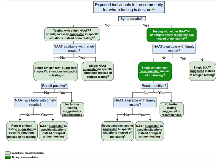
The IDSA also published a guideline regarding serology testing with the following recommendations:95
- “The IDSA panel recommends against using serologic testing to diagnose SARS-CoV-2 infection during the first two weeks following symptom onset (strong recommendation, low certainty of evidence).
- The IDSA panel recommends against using IgG antibodies to provide evidence of COVID-19 in symptomatic patients with a high clinical suspicion and repeatedly negative NAAT (strong recommendation, very low certainty of evidence).
- To assist with the diagnosis of multisystem inflammatory syndrome in children (MIS-C), the IDSA panel recommends using both IgG antibody testing and NAAT to provide evidence of current or recent past COVID-19 (strong recommendation, very low certainty of evidence).
- When evidence of previous SARS-CoV-2 infection is desired, the IDSA panel suggests testing for SARS-CoV-2 IgG, IgG/IgM, or total antibodies three to five weeks after symptom onset and suggests against testing for SARS-CoV-2 IgM (conditional recommendation, low certainty of evidence).
- When evidence of prior SARS-CoV-2 infection is desired, the IDSA panel suggests using serologic assays that target nucleocapsid protein rather than spike protein (conditional recommendation, low certainty of evidence).
- In individuals with previous SARS-CoV-2 infection or vaccination, the IDSA panel suggests against routine serologic testing given no demonstrated benefit to improving patient outcomes (conditional recommendation, very low certainty of evidence).”95
Infectious Diseases Society of America (IDSA)/American Society for Microbiology (ASM)
In 2022, IDSA and ASM released a consensus review document on the clinical and infection prevention applications for SARS-CoV-2 genotyping. In it, they cover clinical use cases for genotyping, methods of genotyping, assay validation and regulatory requirements, clinical reporting for laboratories, and emerging issues in clinical SARS-CoV-2 sequencing. Overall, they report that “while clinical uses of SARS-CoV2 genotyping are currently limited, rapid technological change along with a growing ability to interpret variants in real time foretell a growing role for SARS-CoV-2 genotyping in clinical care as continuing data emerge on vaccine and therapeutic efficacy.”96
Society for Healthcare Epidemiology of America (SHEA)/American Society of Anesthesiologists (ASA)/Anesthesia Patient Safety Foundation (APSF)
In late 2022, SHEA published recommendations on screening for SARS-CoV-2 in an asymptomatic population. Here, they note that testing of asymptomatic patients was an attempt to reduce the risk of nosocomial transmission but has been an extensive and resource intensive process with unclear benefit when added to other layers of infection prevention mitigation controls. They also note that “the logistic challenges and costs related to screening program implementation, data noting the lack of substantial aerosol generation with elective controlled intubation, extubation, and other procedures, and the adverse patient and facility consequences of asymptomatic screening call into question the utility of this infection prevention intervention.” Based on their findings, SHEA “recommends against routine universal use of asymptomatic screening for SARS-CoV-2 in healthcare facilities. Specifically, preprocedure asymptomatic screening is unlikely to provide incremental benefit in preventing SARS-CoV-2 transmission in the procedural and perioperative environment when other infection prevention strategies are in place, and it should not be considered a requirement for all patients. Admission screening may be beneficial during times of increased virus transmission in some settings where other layers of controls are limited (eg, behavioral health, congregate care, or shared patient rooms), but widespread routine use of admission asymptomatic screening is not recommended over strengthening other infection prevention controls.”97
This statement is supported by the ASA and the APSF. They specifically note that the “SHEA recommendations provide a rationale for considering a move away from universal screening. Such a change considers the potential adverse consequences of testing for SARS-CoV-2 in asymptomatic patients. Moreover, we recommend that each facility develop a risk/benefit analysis that includes local/facility infection prevention assessment (e.g., patient population, facility physical layout, and community incidence and transmission of COVID-19 as defined in the SHEA Board Commentary), and a robust system of controls and interventions to prevent virus transmission (“Swiss Cheese” model). The recommendations by SHEA should be considered along with these updated recommendations to operationalize a robust and safe perioperative screening and targeted testing program for the benefit of our patients, our healthcare workers, other hospital patients and the public.”98
American Association for Clinical Chemistry (AACC)
The AACC released a set of recommendations for “implementing and interpreting SARS-CoV-2 EUA and LDT serologic testing in clinical laboratories.” Serologic testing is currently only used for serum, plasma, and “less frequently, whole-blood or dried blood spots,” but not for other sample types, like saliva and cerebrospinal fluid. Serologic testing is “not recommended as the primary approach for diagnosis of SARS-CoV-2 infection.” For the recommended use of serologic testing, the AACC stated the following:
- “Serologic testing may be offered as an approach to support diagnosis of COVID -19 illness in symptomatic patients and late phase negative molecular testing or for patients presenting with late complications such as multisystem inflammatory syndrome in children (MIS -C).
- Serologic testing can help identify people who may have been infected with or have recovered from the SARS -CoV -2 infection.
- Serologic testing can be used to screen potential convalescent plasma donors and in the manufacture of convalescent plasma.
- Serologic testing can be used for epidemiology and seroprevalence studies.
- Serologic testing can be used for vaccine response and efficacy studies.”
Regarding serologic testing limitations, the AACC stated the following:
- “False positive results may occur.
- Negative results do not preclude acute SARS-CoV-2 infection or viral shedding.
- Serologic tests may not differentiate between natural infection and vaccine response.
- Serologic results should not be used for
- Determining individual protective immunity
- Return to work decisions
- Cohorting individuals in congregate settings
- Assessment of convalescent plasma recipients
- Use of Personal Protective Equipment
- Placement of high risk job functions.”99
European Centre for Disease Prevention and Control (ECDC)
The ECDC in their guidance for laboratory support in the EU/EEA recommends using WHO-recommended testing strategies for the diagnosis and confirmation of COVID-19.100
In the ECDC’s guideline titled “COVID-19 testing strategies and objectives”, the ECDC recommends performing laboratory testing in accordance with the WHO case definition. The following populations should be tested:101
- “Ideally, all people with COVID-19 symptoms should be tested as soon as possible after symptom onset. This requires easy access to testing for all, including non-residents. Test result turnaround time should be minimized, people testing positive should isolate and timely contact tracing should be carried out, ensuring that all close contacts are tested, irrespective of symptoms.
- All patients with acute respiratory symptoms in hospitals and in other healthcare settings, and all specimens from sentinel primary care surveillance should be tested for both SARS-CoV-2 and influenza during the influenza season to monitor incidence and trends over time.
- Healthcare and social care settings require intensive testing when there is documented community transmission. Periodic and comprehensive testing of all staff and residents/patients is recommended to prevent nosocomial transmission. Furthermore, all patients/residents should be tested upon or just prior to admission.
- Clusters or outbreaks may occur in certain settings, such as workplaces, educational facilities, prisons, and migrant detention centres. Testing policies and systems should be in place for rapid detection and control to protect the relevant populations in these settings and to protect the community from amplified transmission.
- Countries experiencing high SARS-CoV-2 transmission in a local community should consider testing the whole population of the affected area. This would enable identification of infectious COVID-19 cases and allow for their prompt isolation to interrupt chains of transmission. Depending on the epidemiological situation, size and population density of the affected area, such an approach could be less disruptive for society than having to introduce and ensure compliance with more stringent public health measures.
- To prevent re-introduction, countries or subnational areas that achieved sustained control of the circulation of SARS-CoV-2 should, in addition to quarantine measures, consider targeted testing and follow-up of individuals coming from other areas within the same country, or from other countries that have not yet achieved sustained control of the virus.”101
The ECDC notes that “Genomic surveillance of SARS-CoV-2 is essential to detect, monitor and assess virus variants that can result in increased transmissibility, disease severity, or have other adverse effects on public health and social control measures. Obtaining timely and accurate information on the emergence and circulation of variants of concern (VOCs) and variants of interest (VOIs) requires robust surveillance systems, including integrated genome sequencing with a well-defined sampling and sequencing strategy to ensure representativeness and reliability of findings.”101,102
The EDCD released guidelines on the use of antibody tests for SARS-CoV-2 in 2022. The key messages are:
- “At present, antibody tests are mostly used in research studies (mainly sero-epidemiological) at population level rather than for individual diagnosis of COVID-19 cases.
- A positive antibody test result can indicate a previous infection or vaccination but cannot be used to determine whether an individual is currently infectious or protected against infection.
- In the absence of a positive diagnostic test result, antibody tests cannot determine the time of infection.
- The antibody titres that correlate with protection from infection are currently unknown.
- There are a variety of antibody tests available and it is extremely difficult to compare their results due to the diversity and lack of standardisation.
- Antibody tests that target the spike protein are unable to distinguish between those who have been previously infected and those who have received at least one dose of a SARS-CoV-2 vaccine.
- There is a risk that the antibodies detected by the commercial tests currently in use will not prevent infection with newly emerging SARS-CoV-2 variants.”103
Pediatric Infectious Diseases Society (PIDS)
The Pediatric Infectious Diseases Society (PIDS) Pediatric COVID-19 Therapies Taskforce has released updated guidance on the management of COVID-19 in children and adolescents. They note that most pediatric infections are mild to moderate; however, a subset of patients are at increased risk for severe or critical illness, as well as potential long-term complications.104
“In children and adolescents with COVID-19 infection, the panel suggests using risk stratification that takes into consideration the assessment of pre-existing conditions, exacerbating factors, and prior immunity to determine appropriate management.
- Children and adolescents may be considered at high risk if all of the following conditions are met:
- They have a definite or probable risk factor for severe COVID-19. Severe immunocompromise, obesity, diabetes, prematurity, and chronic cardiac, neurologic (seizures), or pulmonary disease (excluding asthma) can be considered definite risk factors. Probable risk factors include sickle cell disease, mild/moderate immunocompromise, neuro-disabilities (trisomy 21), and chronic kidney, gastrointestinal, and liver disease.
- They have an exacerbating condition, including multiple (≥2) comorbidities, a severe or poorly controlled comorbidity, or being <1 or ≥12 years of age.
- They have no prior immunity, defined as up-to-date immunization or recent infection (within the previous 4 months) in an immunocompetent host.
- Children and adolescents may be considered at moderate risk if they have definite or probable risk factors for severe COVID-19 but no exacerbating conditions or if they are immunocompetent and have prior immunity.
- Children and adolescents with no risk factors for severe COVID-19 may be considered at low risk for severe COVID-19.”104
Regarding treatment of COVID-19 in pediatric patients PIDS recommends:
- “For pediatric patients with SARS-CoV-2 infection who do not require hospitalization for COVID-19 and are at low risk for progression to severe disease, the panel suggests against specific treatment for SARS-CoV-2.
- For pediatric patients with SARS-CoV-2 infection who do not require hospitalization for COVID-19 and are at moderate risk for progression to severe disease, consider specific treatment for SARS-CoV-2 on a case-by-case basis.
- For pediatric patients with SARS-CoV-2 infection who do not require hospitalization for COVID-19 and are at high risk for progression to severe disease, the panel suggests administering specific treatment for SARS-CoV-2.
- For pediatric patients with SARS-CoV-2 infection who do not require hospitalization for COVID-19 and for whom specific treatment is considered or suggested, the panel suggests treatment with one of the following:
- ritonavir-boosted nirmatrelvir, remdesivir, or a mAb (if expected to be effective against circulating strains).
- If none of these options is available or clinically appropriate, consider treatment with molnupiravir for patients ≥18 years of age.”104
American College of Rheumatology (ACR)
The ACR published guidance regarding MIS-C associated with COVID-19. In it, they list SARS-CoV-2 IgG, IgM, and IgA as part of the diagnostic pathway for MIS-C.105
In a December 5, 2020 update of the above guidelines, the ACR states that ESR, CRP, and testing for SARS-CoV-2 (by PCR or serology) should be considered a “tier 1” (first-line evaluation) for MIS-C.106
In a February 3, 2022 update of the above guideline, the ACR added new information concerning immunomodulatory treatment in MIS-C, hyperinflammation in COVID-19, as well as statements on thrombotic risk and anticoagulation in MIS-C.107
References
1. CDC. About COVID-19. Updated June 13, 2024. https://www.cdc.gov/covid/about/index.html
2. CDC. Symptoms of COVID-19. Updated March 10, 2025. https://www.cdc.gov/covid/signs-symptoms/
3. CDC. Multisystem Inflammatory Syndrome: Case Definitions and Reporting. Updated Febuary 19, 2026. https://www.cdc.gov/mis/hcp/case-definition-reporting/index.html
4. Cucinotta D, Vanelli M. WHO Declares COVID-19 a Pandemic. Acta Biomed. Mar 19 2020;91(1):157-160. doi:10.23750/abm.v91i1.9397
5. WHO. SARS (Severe Acute Respiratory Syndrome). https://www.who.int/ith/diseases/sars/en/
6. WHO. Middle East respiratory syndrome coronavirus (MERS-CoV). https://www.who.int/emergencies/mers-cov/en/
7. WHO. COVID-19 Global Risk Assessment. Updated February 6, 2025. https://www.who.int/publications/m/item/covid-19-global-risk-assessment
8. Gandhi R. COVID-19: Clinical features. Updated May 7, 2025. https://www.uptodate.com/contents/covid-19-clinical-features
9. Yang X, Yu Y, Xu J, et al. Clinical course and outcomes of critically ill patients with SARS-CoV-2 pneumonia in Wuhan, China: a single-centered, retrospective, observational study. Lancet Respir Med. May 2020;8(5):475-481. doi:10.1016/s2213-2600(20)30079-5
10. WHO. COVID-19 Clinical management: living guidance. Updated November 23, 2021. https://apps.who.int/iris/bitstream/handle/10665/349321/WHO-2019-nCoV-clinical-2021.2-eng.pdf
11. Kawasuji H, Takegoshi Y, Kaneda M, et al. Transmissibility of COVID-19 depends on the viral load around onset in adult and symptomatic patients. PLOS ONE. 2020;15(12):e0243597. doi:10.1371/journal.pone.0243597
12. Cevik M, Tate M, Lloyd O, Maraolo AE, Schafers J, Ho A. SARS-CoV-2, SARS-CoV, and MERS-CoV viral load dynamics, duration of viral shedding, and infectiousness: a systematic review and meta-analysis. The Lancet Microbe. January 1 2021;2(1):E13-E22. doi:10.1016/S2666-5247(20)30172-5
13. Griffin D. Viral Load as a Predictor of COVID-19 Patient Outcomes. Updated December 31, 2020. https://www.cuimc.columbia.edu/news/viral-load-predictor-covid-19-patient-outcomes
14. DeBiasi RL, Song X, Delaney M, et al. Severe COVID-19 in Children and Young Adults in the Washington, DC Metropolitan Region. J Pediatr. May 13 2020;doi:10.1016/j.jpeds.2020.05.007
15. WHO. Multisystem inflammatory syndrome in children and adolescents with COVID-19. Updated May 15, 2020. https://www.who.int/publications-detail/multisystem-inflammatory-syndrome-in-children-and-adolescents-with-covid-19
16. Verdoni L, Mazza A, Gervasoni A, et al. An outbreak of severe Kawasaki-like disease at the Italian epicentre of the SARS-CoV-2 epidemic: an observational cohort study. Lancet. May 13 2020;doi:10.1016/s0140-6736(20)31103-x
17. Jones VG, Mills M, Suarez D, et al. COVID-19 and Kawasaki Disease: Novel Virus and Novel Case. Hosp Pediatr. Apr 7 2020;doi:10.1542/hpeds.2020-0123
18. Baum SG. Adult Multisystem Inflammatory Syndrome Associated with COVID-19. Updated October 21, 2020. https://www.jwatch.org/na52622/2020/10/21/adult-multisystem-inflammatory-syndrome-associated-with
19. Morris SB, Schwartz NG, Patel P, et al. Case Series of Multisystem Inflammatory Syndrome in Adults Associated with SARS-CoV-2 Infection - United Kingdom and United States, March-August 2020. MMWR Morb Mortal Wkly Rep. Oct 9 2020;69(40):1450-1456. doi:10.15585/mmwr.mm6940e1
20. CDC. SARS-CoV-2 Variant Classications and Denitions. Updated December 1, 2021. https://stacks.cdc.gov/view/cdc/112177
21. CDC. Variants and Genomic Surveillance. Updated August 28, 2025. https://www.cdc.gov/covid/php/variants/variants-and-genomic-surveillance.html
22. CDC. Interim Clinical Considerations for Use of COVID-19 Vaccines Currently Approved or Authorized in the United States. Updated November 4, 2025. https://www.cdc.gov/vaccines/covid-19/clinical-considerations/interim-considerations-us.html
23. Corman VM, Lienau J, Witzenrath M. [Coronaviruses as the cause of respiratory infections]. Internist (Berl). Nov 2019;60(11):1136-1145. Coronaviren als Ursache respiratorischer Infektionen. doi:10.1007/s00108-019-00671-5
24. Ludwig S, Zarbock A. Coronaviruses and SARS-CoV-2: A Brief Overview. Anesth Analg. Mar 31 2020;doi:10.1213/ane.0000000000004845
25. CDC. Overview of Testing for SARS-CoV-2. Updated August 29, 2024. https://www.cdc.gov/covid/hcp/clinical-care/overview-testing-sars-cov-2.html
26. Veroniki AA, Tricco AC, Watt J, et al. Rapid antigen-based and rapid molecular tests for the detection of SARS-CoV-2: a rapid review with network meta-analysis of diagnostic test accuracy studies. BMC Medicine. 2023/03/29 2023;21(1):110. doi:10.1186/s12916-023-02810-0
27. Lippi G, Simundic AM, Plebani M. Potential preanalytical and analytical vulnerabilities in the laboratory diagnosis of coronavirus disease 2019 (COVID-19). Clin Chem Lab Med. Mar 16 2020;doi:10.1515/cclm-2020-0285
28. Chan JF, Yip CC, To KK, et al. Improved Molecular Diagnosis of COVID-19 by the Novel, Highly Sensitive and Specific COVID-19-RdRp/Hel Real-Time Reverse Transcription-PCR Assay Validated In Vitro and with Clinical Specimens. J Clin Microbiol. Apr 23 2020;58(5)doi:10.1128/jcm.00310-20
29. CDC. CDC 2019-Novel Coronavirus (2019-nCoV) Real-Time RT-PCR Diagnostic Panel. Updated March 7, 2023. https://www.fda.gov/media/134922/download
30. FDA. Accelerated Emergency Use Authorization (EUA) Summary. Updated June 21, 2022. https://www.fda.gov/media/136151/download
31. Braeye T, Abrams S, Hens N. Factors influencing SARS-CoV-2 IgG test sensitivity: A Bayesian analysis of seroconversion and seroreversion by time since infection, test, age and disease severity. PLoS One. 2026;21(2):e0328144. doi:10.1371/journal.pone.0328144
32. The Native Antigen Company. Why We Need Antigen and Antibody Tests for COVID-19. The Native Antigen Company. Updated March 24, 2020. https://thenativeantigencompany.com/why-we-need-antigen-and-antibody-tests-for-covid-19/
33. Kaduskar O, Gurav YK, Deshpande K, et al. Understanding the dynamics of IgM & IgG antibodies in COVID-19-positive patients. Indian J Med Res. May-Jun 2022;155(5&6):565-569. doi:10.4103/ijmr.IJMR_675_21
34. Espejo AP, Akgun Y, Al Mana AF, et al. Review of Current Advances in Serologic Testing for COVID-19. Am J Clin Pathol. Aug 5 2020;154(3):293-304. doi:10.1093/ajcp/aqaa112
35. Nayagam SN, Coote W, Carroll MTC, et al. Differentiating Severe Acute Respiratory Syndrome Coronavirus 2 Antibody Responses Between Infection and Vaccination: Challenges for Epidemiological Research. The Journal of Infectious Diseases. 2025;232(1):9-13. doi:10.1093/infdis/jiaf295
36. FDA. Policy for Coronavirus Disease-2019 Tests During the Public Health Emergency (Revised). FDA. Updated January 2023. https://www.fda.gov/regulatory-information/search-fda-guidance-documents/policy-coronavirus-disease-2019-tests-during-public-health-emergency-revised
37. Zhao J, Yuan Q, Wang H, et al. Antibody responses to SARS-CoV-2 in patients of novel coronavirus disease 2019. Clinical Infectious Diseases. 2020;doi:10.1093/cid/ciaa344
38. Li Q, Chen L, Li F, He A. Long-term evaluation of the seroprevalence of SARS-CoV-2 IgG and IgM antibodies in recovered patients: a meta-analysis. BMC Infectious Diseases. 2023/07/01 2023;23(1):444. doi:10.1186/s12879-023-08425-3
39. Lisboa Bastos M, Tavaziva G, Abidi SK, et al. Diagnostic accuracy of serological tests for covid-19: systematic review and meta-analysis. Bmj. Jul 1 2020;370:m2516. doi:10.1136/bmj.m2516
40. Kontou PI, Braliou GG, Dimou NL, Nikolopoulos G, Bagos PG. Antibody Tests in Detecting SARS-CoV-2 Infection: A Meta-Analysis. Diagnostics (Basel). May 19 2020;10(5)doi:10.3390/diagnostics10050319
41. Caturegli G, Materi J, Howard BM, Caturegli P. Clinical Validity of Serum Antibodies to SARS-CoV-2 : A Case-Control Study. Ann Intern Med. Oct 20 2020;173(8):614-622. doi:10.7326/m20-2889
42. Fox T, Geppert J, Dinnes J, et al. Antibody tests for identification of current and past infection with SARS-CoV-2. Cochrane Database Syst Rev. Nov 17 2022;11(11):Cd013652. doi:10.1002/14651858.CD013652.pub2
43. NIH. Authorized Tests for COVID-19 and Multiplex Diagnostics. Updated 2026. https://www.nibib.nih.gov/programs/radx-tech-program/authorized-tests
44. BD. BD Receives FDA 510(k) Clearance for Rapid Point-of-Care COVID-19 Test. Updated July 30, 2025. https://investors.bd.com/news-events/press-releases/detail/895/bd-receives-fda-510k-clearance-for-rapid-point-of-care-covid-19-test
45. Diagnostics R. LumiraDx SARS-CoV-2 Ag Test. Updated March 26, 2026. https://diagnostics.roche.com/us/en/c/lumira-sars-cov-2-ag-test.html
46. Quidel Corporation. Sofia 2 SARS Antigen FIA. FDA. https://www.fda.gov/media/137885/download
47. FDA. In Vitro Diagnostics EUAs. Updated November 8, 2023. https://www.fda.gov/medical-devices/coronavirus-disease-2019-covid-19-emergency-use-authorizations-medical-devices/vitro-diagnostics-euas
48. Mathias Weis D, David Ruben Teindl L, Mia E, et al. Preapproval and postapproval diagnostic test accuracy of food and drug administration–authorized rapid antigen SARS-CoV-2 tests used according to instruction: a systematic review and meta-analysis. Clinical Microbiology and Infection. 2025;31(10):1630-1638. doi:10.1016/j.cmi.2025.07.009
49. Seo G, Lee G, Kim MJ, et al. Rapid Detection of COVID-19 Causative Virus (SARS-CoV-2) in Human Nasopharyngeal Swab Specimens Using Field-Effect Transistor-Based Biosensor. ACS Nano. Apr 28 2020;14(4):5135-5142. doi:10.1021/acsnano.0c02823
50. Scohy A, Anantharajah A, Bodéus M, Kabamba-Mukadi B, Verroken A, Rodriguez-Villalobos H. Low performance of rapid antigen detection test as frontline testing for COVID-19 diagnosis. J Clin Virol. Aug 2020;129:104455. doi:10.1016/j.jcv.
51. Mak GC, Cheng PK, Lau SS, et al. Evaluation of rapid antigen test for detection of SARS-CoV-2 virus. J Clin Virol. Aug 2020;129:104500. doi:10.1016/j.jcv.
52. Lambert-Niclot S, Cuffel A, Le Pape S, et al. Evaluation of a Rapid Diagnostic Assay for Detection of SARS-CoV-2 Antigen in Nasopharyngeal Swabs. J Clin Microbiol. Jul 23 2020;58(8)doi:10.1128/jcm.00977-20
53. Soni A, Herbert C, Lin H, et al. Performance of Rapid Antigen Tests to Detect Symptomatic and Asymptomatic SARS-CoV-2 Infection : A Prospective Cohort Study. Ann Intern Med. Jul 2023;176(7):975-982. doi:10.7326/m23-0385
54. Peacock WF, Soto-Ruiz KM, House SL, et al. Utility of COVID-19 antigen testing in the emergency department. Journal of the American College of Emergency Physicians Open. 2022;3(1):e12605. doi:10.1002/emp2.12605
55. FDA. Emergency Use Authorization. Updated December 23, 2024. https://www.fda.gov/emergency-preparedness-and-response/mcm-legal-regulatory-and-policy-framework/emergency-use-authorization
56. BioFire. BIOFIRE® Respiratory 2.1 (RP2.1) Panel. BioMerieux. https://www.biomerieux.com/us/en/our-offer/clinical-products/biofire-respiratory-panels.html
57. Qiagen GmbH. QIAstat-Dx® Respiratory SARS-CoV2 Panel Instructions for Use (Handbook). https://www.fda.gov/media/136571/download
58. GenMark Diagnostics. ePlex Respiratory Pathogen Panel 2. https://www.fda.gov/media/142902/download
59. FDA. Influenza SARS-CoV-2 (Flu SC2) Multiplex Assay. Updated August 5, 2021. https://www.fda.gov/media/139744/download
60. bioMérieux. BIOFIRE® Respiratory 2.1 (RP2.1) Panel with SARS-CoV-2 obtains De Novo FDA Authorization. Updated March 18, 2021. https://www.biomerieux.com/us/en/journalists/press-releases/biofirer-respiratory-21-rp21-panel-sars-cov-2-obtains-de-novo-fda-authorization.html
61. Qiagen. QIAstat-Dx Respiratory SARS-CoV-2 Panel - Canada. Updated 2026. https://www.qiagen.com/ca/products/diagnostics-and-clinical-research/infectious-disease/qiastat-dx-syndromic-testing/qiastat-dx-ca#product-details
62. Diagnostics R. cobas® eplex respiratory pathogen panel 2. Updated July 2026. https://diagnostics.roche.com/global/en/products/lab/cobas-eplex-rp-panel-2-pid00000004.html
63. CDC. CDC’s Influenza SARS-CoV-2 Multiplex Assay. Updated September 2, 2025. https://www.cdc.gov/flu/php/laboratories/influenza-sars-cov-2-multiplex-assay.html
64. FDA. CDC Influenza SARS-CoV-2 (Flu SC2) Multiplex Assay. Updated August 6, 2024. https://www.fda.gov/media/139743/download
65. Poljak M, Korva M, Gašper NK, et al. Clinical Evaluation of the cobas SARS-CoV-2 Test and a Diagnostic Platform Switch during 48 Hours in the Midst of the COVID-19 Pandemic. Journal of Clinical Microbiology. 2020;58(6):e00599-20. doi:doi:10.1128/JCM.00599-20
66. designs B. BIOSYNEX AMPLIQUICK® Respiratory Triplex. Updated 2026. https://www.biosynex.com/fiches-produit/biologiste-medecin/biologie-moleculaire/ampliquick/virologie/triplex-respiratoire/
67. Mboumba Bouassa R-S, Tonen-Wolyec S, Veyer D, Péré H, Bélec L. Analytical performances of the AMPLIQUICK® Respiratory Triplex assay for simultaneous detection and differentiation of SARS-CoV-2, influenza A/B and respiratory syncytial viruses in respiratory specimens. PloS one. 2022;17(1):e0262258. doi:10.1371/journal.pone.0262258
68. Dao Thi VL, Herbst K, Boerner K, et al. A colorimetric RT-LAMP assay and LAMP-sequencing for detecting SARS-CoV-2 RNA in clinical samples. Science Translational Medicine. 2020;12(556):eabc7075. doi:10.1126/scitranslmed.abc7075
69. Nagura-Ikeda M, Imai K, Tabata S, et al. Clinical evaluation of self-collected saliva by RT-qPCR, direct RT-qPCR, RT-LAMP, and a rapid antigen test to diagnose COVID-19. J Clin Microbiol. Jul 7 2020;doi:10.1128/jcm.01438-20
70. Wang R, Qian C, Pang Y, et al. opvCRISPR: One-pot visual RT-LAMP-CRISPR platform for SARS-cov-2 detection. Biosensors and Bioelectronics. 10/26/2020 2020;172:112766. doi:10.1016/j.bios.
71. Hulick P. Next-generation DNA sequencing (NGS): Principles and clinical applications. Wolters Kluwer. Updated January 21, 2026. https://www.uptodate.com/contents/next-generation-dna-sequencing-ngs-principles-and-clinical-applications
72. Helix. Helix COVID-19 NGS Test. https://www.fda.gov/media/140917/download
73. FDA. Illumina COVIDSeq Test. Updated April 22, 2021. https://www.fda.gov/media/138778/download
74. Ahmadi Z, Maleki A, Eybpoosh S, et al. Comparison of a Multiplex Real-Time PCR Technique with Oxford Nanopore Technologies Next-Generation Sequencing for Identification of SARS-CoV-2 Variants of Concern. Intervirology. 2023;66(1):136-141. doi:10.1159/000534067
75. Oude Munnink BB, Nieuwenhuijse DF, Stein M, et al. Rapid SARS-CoV-2 whole-genome sequencing and analysis for informed public health decision-making in the Netherlands. Nature Medicine. 2020/09/01 2020;26(9):1405-1410. doi:10.1038/s41591-020-0997-y
76. CDC. About Whole Genome Sequencing. Updated January 8, 2024. https://www.cdc.gov/pulsenet/php/wgs/
77. Sri Santosh T, Parmar R, Anand H, Srikanth K, Saritha M. A Review of Salivary Diagnostics and Its Potential Implication in Detection of Covid-19. Cureus. Apr 17 2020;12(4):e7708. doi:10.7759/cureus.7708
78. To KKW, Yip CCY, Lai CYW, et al. Saliva as a diagnostic specimen for testing respiratory virus by a point-of-care molecular assay: a diagnostic validity study. Clin Microbiol Infect. Mar 2019;25(3):372-378. doi:10.1016/j.cmi.2018.06.009
79. FDA. Accelerated Emergency Use Authorization (EUA) Summary SARS-CoV-2 RT-PCR Assay. https://www.fda.gov/media/141192/download
80. Pooled Testing and Its Applications in the COVID-19 Pandemic , bookTitle= Pandemics: Insurance and Social Protection. Springer International Publishing; 2022:217--249.
81. Yelin I, Aharony N, Shaer Tamar E, et al. Evaluation of COVID-19 RT-qPCR test in multi-sample pools. Clin Infect Dis. May 2 2020;doi:10.1093/cid/ciaa531
82. Hogan CA, Sahoo MK, Pinsky BA. Sample Pooling as a Strategy to Detect Community Transmission of SARS-CoV-2. Jama. Apr 6 2020;323(19):1967-9. doi:10.1001/jama.2020.5445
83. WHO. Diagnostic testing for SARS-CoV-2. Updated September 11, 2020. https://www.who.int/publications/i/item/diagnostic-testing-for-sars-cov-2
84. WHO. "Immunity passports" in the context of COVID-19. Updated April 24, 2020. https://www.who.int/news-room/commentaries/detail/immunity-passports-in-the-context-of-covid-19
85. WHO. COVID-19 natural immunity. Updated May 10, 2021. https://iris.who.int/bitstream/handle/10665/341241/WHO-2019-nCoV-Sci-Brief-Natural-immunity-2021.1-eng.pdf
86. WHO. Use of SARS-CoV-2 antigen-detection rapid diagnostic tests for COVID-19 self-testing. Updated March 9, 2022. https://www.who.int/publications/i/item/WHO-2019-nCoV-Ag-RDTs-Self_testing-2022.1
87. WHO. WHO policy brief: COVID-19 testing. Updated December 10, 2024. https://www.who.int/publications/m/item/who-policy-brief-covid-19-testing
88. WHO. Antigen-detection in the diagnosis of SARS-CoV-2 infection. Updated Septemeber 11, 2021. https://www.who.int/publications/i/item/antigen-detection-in-the-diagnosis-of-sars-cov-2infection-using-rapid-immunoassays
89. CDC. Testing for COVID-19. Updated March 10, 2025. https://www.cdc.gov/covid/testing/index.html
90. CDC. Information for Pediatric Healthcare Providers. Updated Febuary 05, 2026. https://www.cdc.gov/covid/hcp/clinical-care/for-pediatric-hcp.html
91. CDC. People with Certain Medical Conditions and COVID-19 Risk Factors. Updated June 11, 2025. https://www.cdc.gov/covid/risk-factors/index.html
92. CDC. Long COVID Basics. Updated March 9, 2026. https://www.cdc.gov/covid/long-term-effects/
93. AMA. Serological testing for SARS-CoV-2 antibodies. American Medical Association. Updated May 14, 2020. https://www.ama-assn.org/delivering-care/public-health/serological-testing-sars-cov-2-antibodies
94. IDSA. Infectious Diseases Society of America Guidelines on the Diagnosis of COVID-19: Molecular Diagnostic Testing. Updated September 6, 2023. https://www.idsociety.org/practice-guideline/covid-19-guideline-diagnostics/
95. IDSA. Infectious Diseases Society of America Guidelines on the Diagnosis of COVID-19: Serologic Testing. Updated February 9, 2024. https://www.idsociety.org/practice-guideline/covid-19-guideline-serology/
96. Greninger AL, Dien Bard J, Colgrove RC, et al. Clinical and Infection Prevention Applications of Severe Acute Respiratory Syndrome Coronavirus 2 Genotyping: An Infectious Diseases Society of America/American Society for Microbiology Consensus Review Document. Clin Infect Dis. Apr 28 2022;74(8):1496-1502. doi:10.1093/cid/ciab761
97. Talbot TR, Hayden MK, Yokoe DS, et al. Asymptomatic screening for severe acute respiratory coronavirus virus 2 (SARS-CoV-2) as an infection prevention measure in healthcare facilities: Challenges and considerations. Infect Control Hosp Epidemiol. Jan 2023;44(1):2-7. doi:10.1017/ice.2022.295
98. ASA, APSF. ASA and APSF Updated Statement on Perioperative Testing for SARS-CoV-2 in the Asymptomatic Patient. Updated December 21, 2022. https://www.apsf.org/news-updates/asa-and-apsf-updated-statement-on-perioperative-testing-for-sars-cov-2-in-the-asymptomatic-patient/
99. Zhang YV, Wiencek J, Meng QH, et al. AACC Practical Recommendations for Implementing and Interpreting SARS-CoV-2 EUA and LDT Serologic Testing in Clinical Laboratories. Clinical Chemistry. 2021;doi:10.1093/clinchem/hvab051
100. ECDC. Diagnostic testing and screening for SARS-CoV-2. European Centre for Disease Prevention and Control. Updated May 30, 2023. https://www.ecdc.europa.eu/en/covid-19/latest-evidence/diagnostic-testing
101. ECDC. Testing strategies for SARS-CoV-2. Updated December 15, 2022. https://www.ecdc.europa.eu/en/covid-19/surveillance/testing-strategies
102. ECDC. Guidance for representative and targeted genomic SARS-CoV-2 monitoring. Updated May 3, 2021. https://www.ecdc.europa.eu/en/publications-data/guidance-representative-and-targeted-genomic-sars-cov-2-monitoring
103. ECDC. Considerations for the use of antibody tests for SARS-CoV-2 – first update. https://www.ecdc.europa.eu/en/publications-data/use-antibody-tests-sars-cov-2
104. Willis ZI, Oliveira CR, Abzug MJ, et al. Guidance for prevention and management of COVID-19 in children and adolescents: A consensus statement from the Pediatric Infectious Diseases Society Pediatric COVID-19 Therapies Taskforce. J Pediatric Infect Dis Soc. Mar 19 2024;13(3):159-185. doi:10.1093/jpids/piad116
105. Henderson LA, Canna SW, Friedman KG, et al. American College of Rheumatology Clinical Guidance for Multisystem Inflammatory Syndrome in Children Associated With SARS-CoV-2 and Hyperinflammation in Pediatric COVID-19: Version 1. Arthritis Rheumatol. Jul 23 2020;doi:10.1002/art.41454
106. Henderson LA, Canna SW, Friedman KG, et al. American College of Rheumatology Clinical Guidance for Pediatric Patients with Multisystem Inflammatory Syndrome in Children (MIS-C) Associated with SARS-CoV-2 and Hyperinflammation in COVID-19. Version 2. Arthritis Rheumatol. Dec 5 2020;doi:10.1002/art.41616
107. Henderson LA, Canna SW, Friedman KG, et al. American College of Rheumatology Clinical Guidance for Multisystem Inflammatory Syndrome in Children Associated With SARS–CoV-2 and Hyperinflammation in Pediatric COVID-19: Version 3. Arthritis & Rheumatology. 2022;74(4):e1-e20. doi:10.1002/art.42062
108. FDA. Transition Plan for Medical Devices That Fall Within Enforcement Policies Issued During the Coronavirus Disease 2019 (COVID-19) Public Health Emergency. Updated March 2023. https://www.fda.gov/regulatory-information/search-fda-guidance-documents/transition-plan-medical-devices-fall-within-enforcement-policies-issued-during-coronavirus-disease
109. FDA. Transition Plan for Medical Devices Issued Emergency Use Authorizations (EUAs) Related to Coronavirus Disease 2019 (COVID-19). Updated March 2023. https://www.fda.gov/regulatory-information/search-fda-guidance-documents/transition-plan-medical-devices-issued-emergency-use-authorizations-euas-related-coronavirus-disease
Coding Section
| CPT |
Code Description |
| 86328 |
Immunoassay for infectious agent antibody(ies), qualitative or semiquantitative, single step method (e.g., reagent strip); severe acute respiratory syndrome coronavirus 2 (SARS-CoV-2) (Coronavirus disease [COVID-19]) |
| 86408 |
Neutralizing antibody, severe acute respiratory syndrome coronavirus 2 (SARS-CoV-2) (Coronavirus disease [COVID19]); screen |
| 86409 |
Neutralizing antibody, severe acute respiratory syndrome coronavirus 2 (SARS-CoV-2) (Coronavirus disease [COVID19]); titer |
| 86413 |
Severe acute respiratory syndrome coronavirus 2 (SARSCoV-2) (Coronavirus disease [COVID-19]) antibody, quantitative |
| 86769 |
Antibody; severe acute respiratory syndrome coronavirus 2 (SARS-CoV-2) (Coronavirus disease [COVID-19]) |
| 87426 |
Infectious agent antigen detection by immunoassay technique, (e.g., enzyme immunoassay [EIA], enzyme-linked immunosorbent assay [ELISA], immunochemiluminometric assay [IMCA]) qualitative or semiquantitative, multiple-step |
| 87635 |
Infectious agent detection by nucleic acid (DNA or RNA); severe acute respiratory syndrome coronavirus 2 (SARS-CoV-2) (Coronavirus disease [COVID-19]), amplified probe technique |
| 87798 |
Infectious agent detection by nucleic acid (DNA or RNA), not otherwise specified; amplified probe technique, each organism |
| 87811 |
Infectious agent antigen detection by immunoassay with direct optical (i.e., visual) observation; severe acute respiratory syndrome coronavirus 2 (SARS-CoV-2) (Coronavirus disease [COVID-19]) |
| 87913 | Infectious agent genotype analysis by nucleic acid (DNA or RNA); severe acute respiratory syndrome coronavirus 2 (SARS-CoV-2) (coronavirus disease [COVID-19]), mutation identification in targeted region(s) |
| 0224U |
Antibody, severe acute respiratory syndrome |
| 0226U |
Surrogate viral neutralization test (sVNT), severe acute respiratory syndrome coronavirus 2 (SARS-CoV-2) (Coronavirus disease [COVID-19]), ELISA, plasma, serum |
| 0408U |
Infectious agent antigen detection by bulk acoustic wave biosensor immunoassay, severe acute respiratory syndrome coronavirus 2 (SARS-CoV-2) (coronavirus disease [COVID-19]) Proprietary test: Omnia™ SARS-CoV-2 Antigen Test Lab/Manufacturer: Qorvo Biotechnologies |
| U0001 |
CDC Novel Coronavirus (2019-nCoV) Real-Time RT-PCR Diagnostic Panel |
| U0002 |
Non-CDC laboratory test for 2019-nCoV (COVID-19), any method |
Procedure and diagnosis codes on Medical Policy documents are included only as a general reference tool for each policy. They may not be all-inclusive.
This medical policy was developed through consideration of peer-reviewed medical literature generally recognized by the relevant medical community, U.S. FDA approval status, nationally accepted standards of medical practice and accepted standards of medical practice in this community and other nonaffiliated technology evaluation centers, reference to federal regulations, other plan medical policies, and accredited national guidelines.
"Current Procedural Terminology © American Medical Association. All Rights Reserved"
History From 2023 Forward